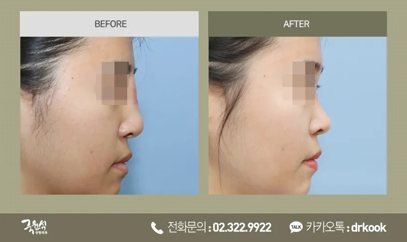

Removal of an Extruded, Infected Implant: Case III
Removal of an Extruded, Infected Implant: Case III
(Removal of infected tissue and protruded silicon implant)
When an implant placed in the nose extrudes, a variety of problems can accompany it.
Once an implant begins to extrude, there is infection and granuloma around the silicone,
the capsule surrounding the silicone thickens, and the once-normal
soft tissue breaks down, leaving the skin thinner.
In some cases the silicone breaks through the thinned skin and becomes exposed on the outside,
and in others it breaks through the mucosa inside the nose.
If an implant has broken through the skin or mucosa, it can be concluded that infection is certainly present inside the nose.
When it has progressed to the point that the implant breaks through the skin, a considerable time has usually passed,
so the implant, granuloma, infection, capsule, scar tissue, and other problems must be removed as soon as possible
in order to prevent and resolve contracture.
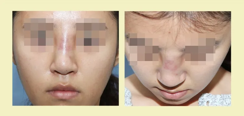
This patient had undergone, 5 years and 4 years earlier at another clinic,
two rhinoplasty procedures.
However, starting 4 months ago, pus and discharge began to come from inside the nose,
and with marked swelling from the tip to the bridge, the patient visited our clinic.
The glabella and dorsum were severely swollen, and accompanying erythema was also noted.
The length of the nose had not changed much, and contracture did not appear to have begun.
On examination of the dorsum and glabella, fluctuance was present,
suggesting that infectious discharge had collected inside the nose.
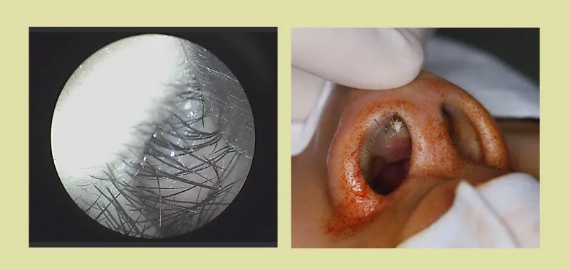
Endoscopic examination of the inside of the nose showed white silicone that had broken through the mucosa
and was exposed over an area of 1.0 X 0.5cm.
The surgical plan was to remove the silicone, capsule, and infected tissue
and thereby treat the infection.
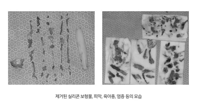
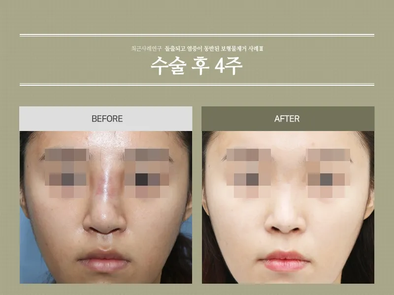
All infected tissue, capsule, silicone, and other material in the subcutaneous layer was removed.
Because the infection had spread widely to the dorsum, tip, and columella, the infected tissue was removed thoroughly.
After complete removal of the infected tissue, the skin had become very thin
and marked skin irregularities remained, but during recovery tissue gradually filled in,
bringing considerable improvement, and the irregularities are expected to fade over time.
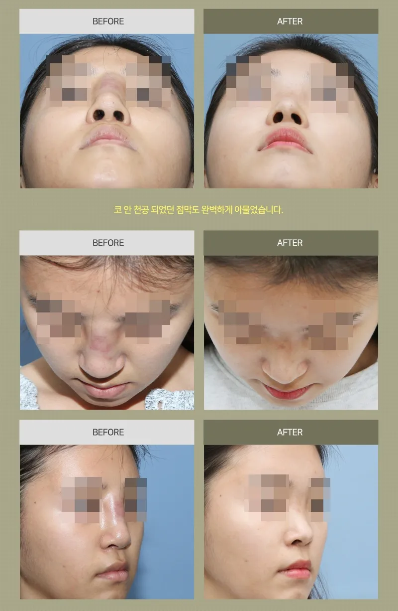
Even during the contraction period, the tip did not turn up,
and the nose returned to its original, natural shape.
The patient wished to have filler treatment later, so it was decided to inject it in 6 months, once the nose has stabilized.