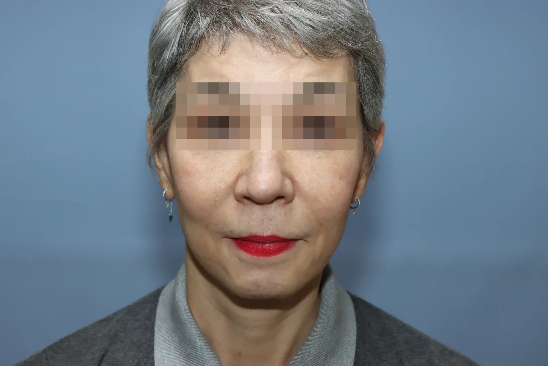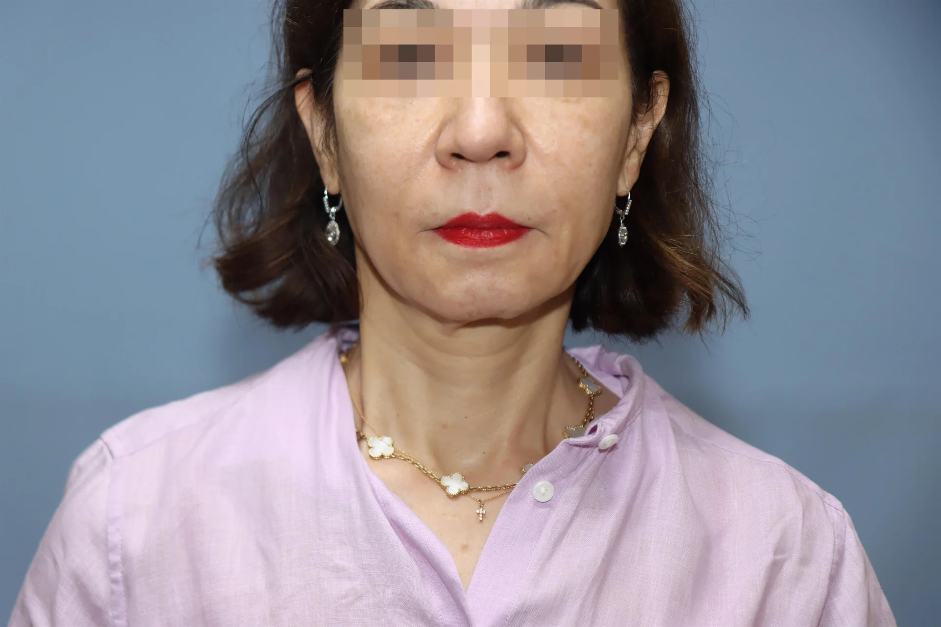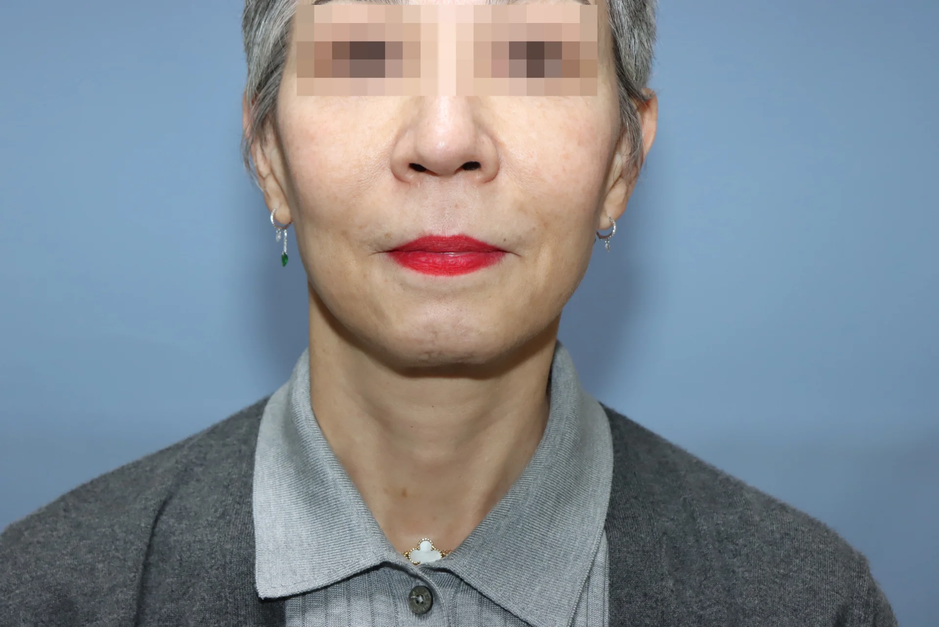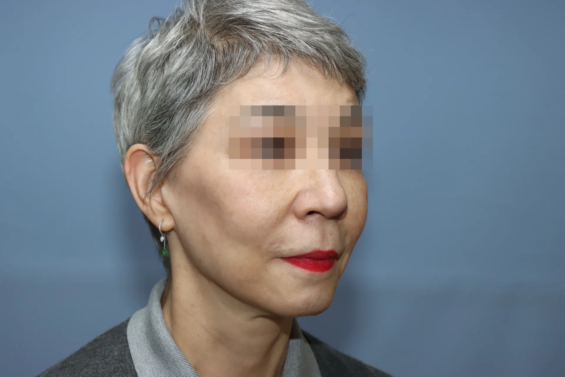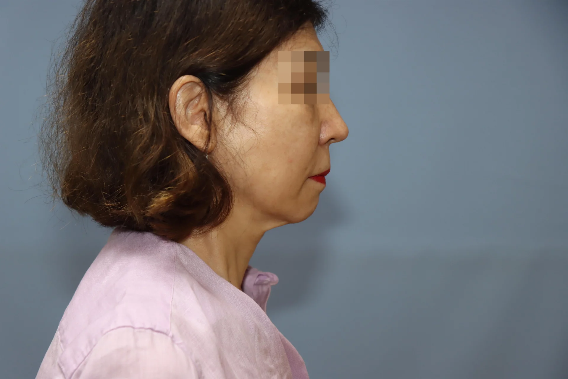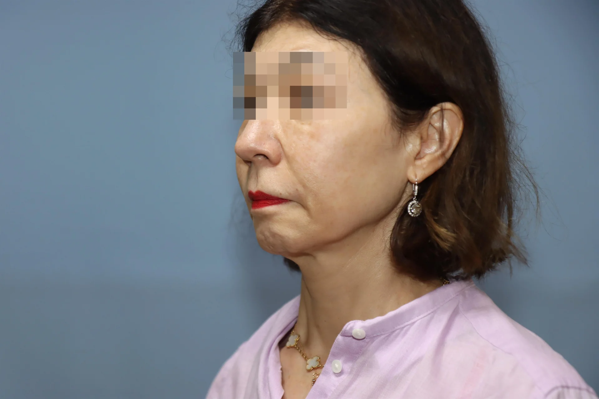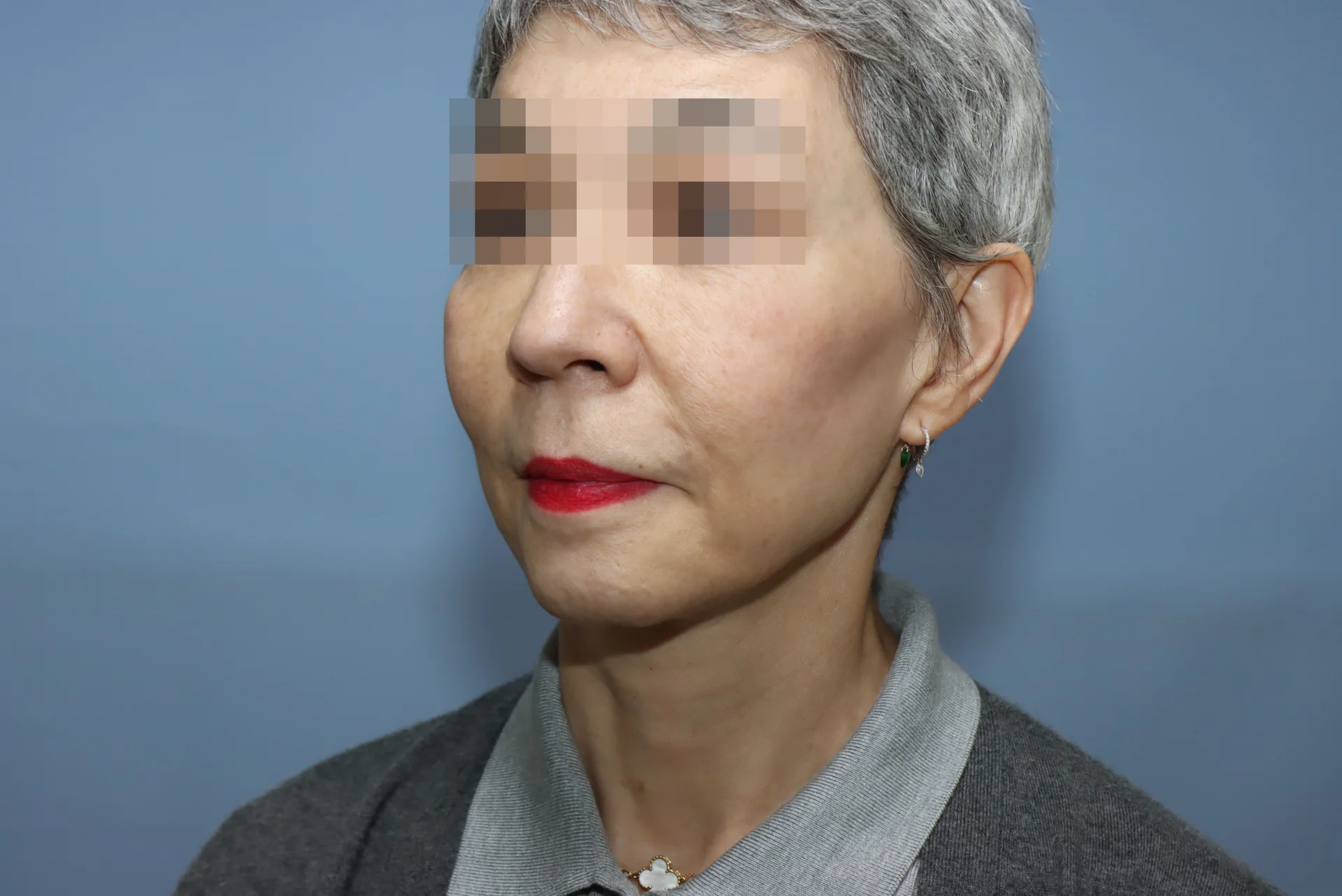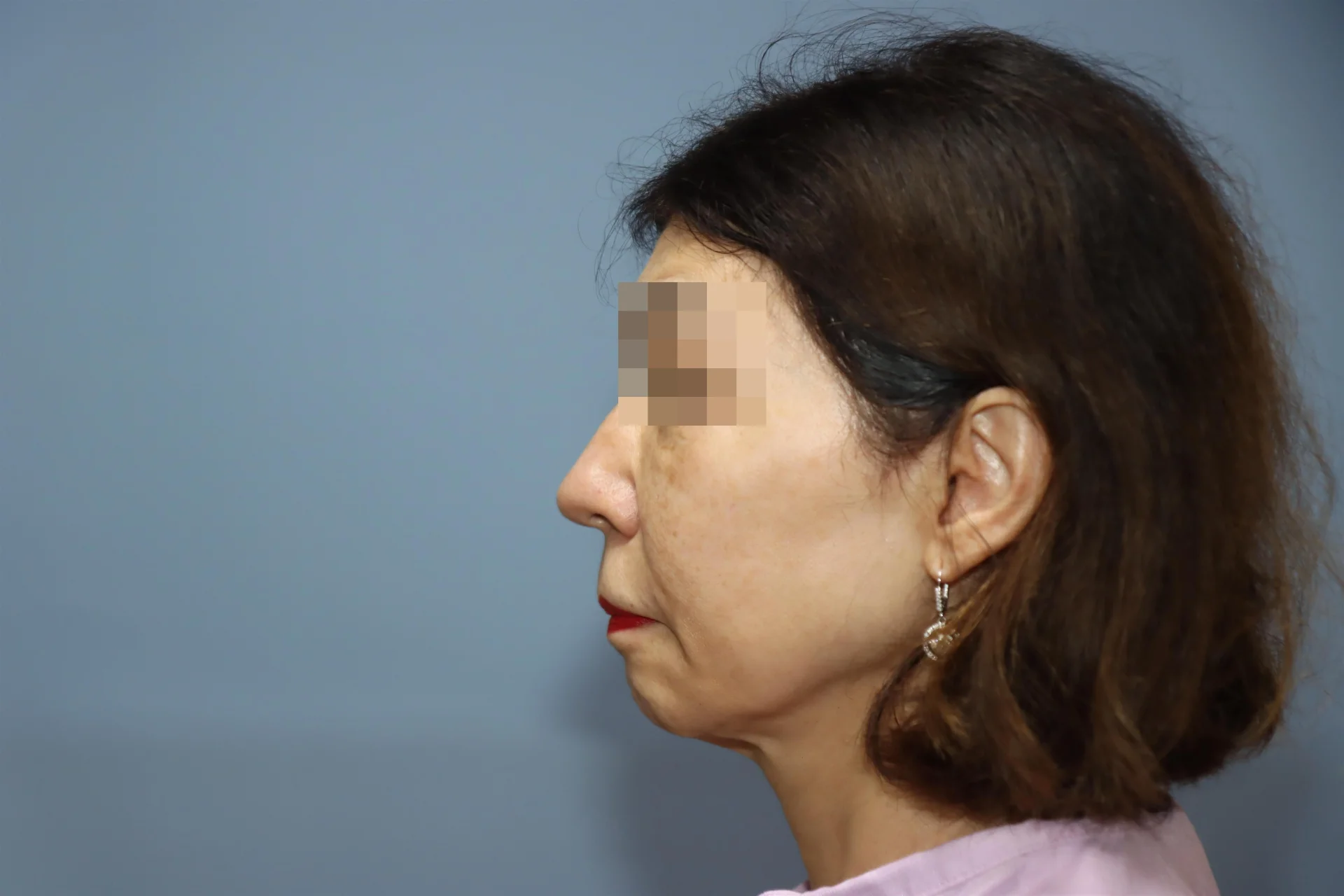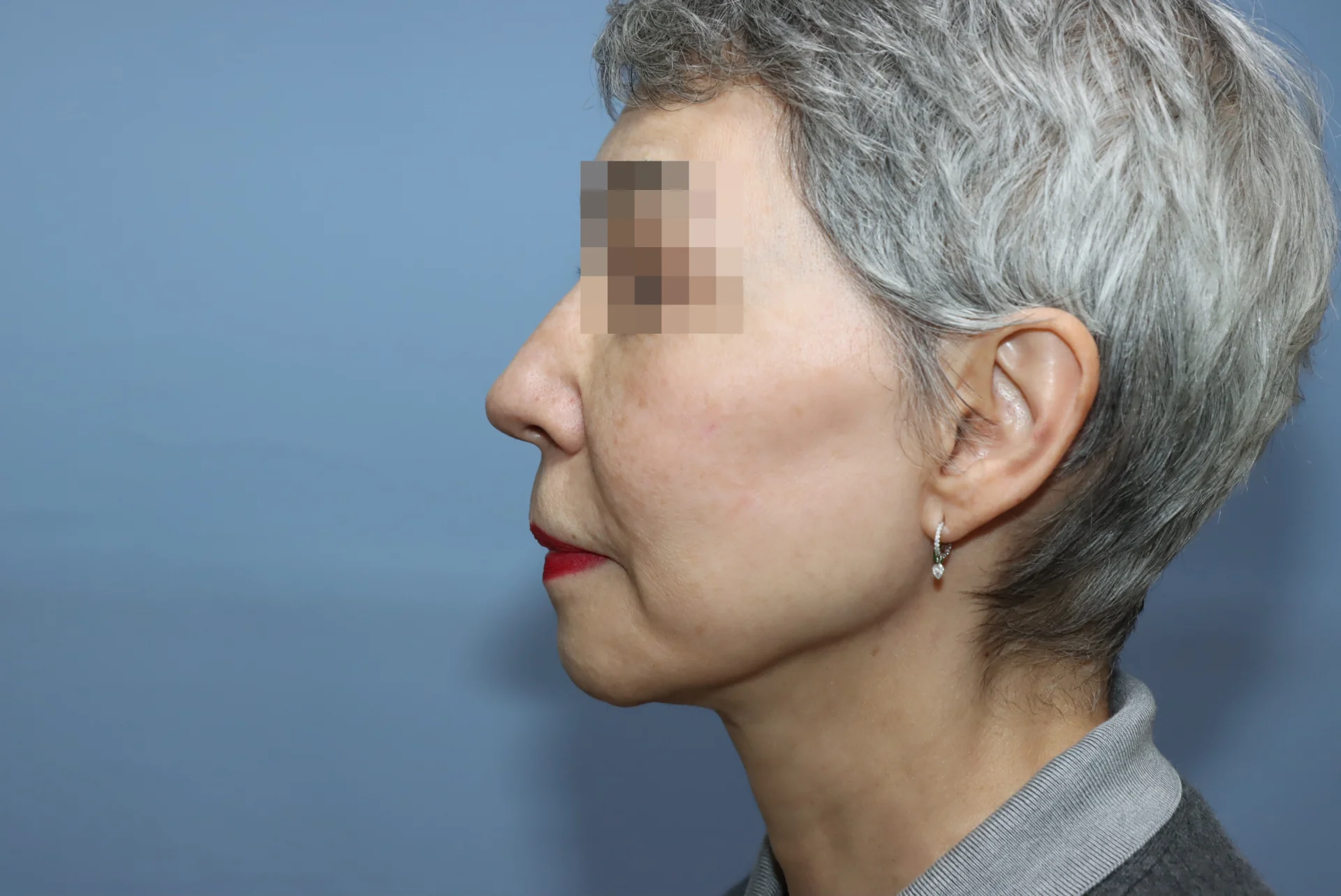

Deep Plane Facelift
Skip to main content

Deep Plane Facelift

Deep Plane Facelift

Deep Plane Facelift

A deep plane facelift is not a surgery that pulls the skin tight;
it repositions the deep soft tissue of the face, which has descended with aging, closer to its original anatomical position.

Method
Anesthesia
Recovery
Medical Review Medical Review: Won-Suk Kook,Jin-Young Kim
Last updated: 2026-10-02

Overview

Deep Plane Facelift

Deep Plane Facelift

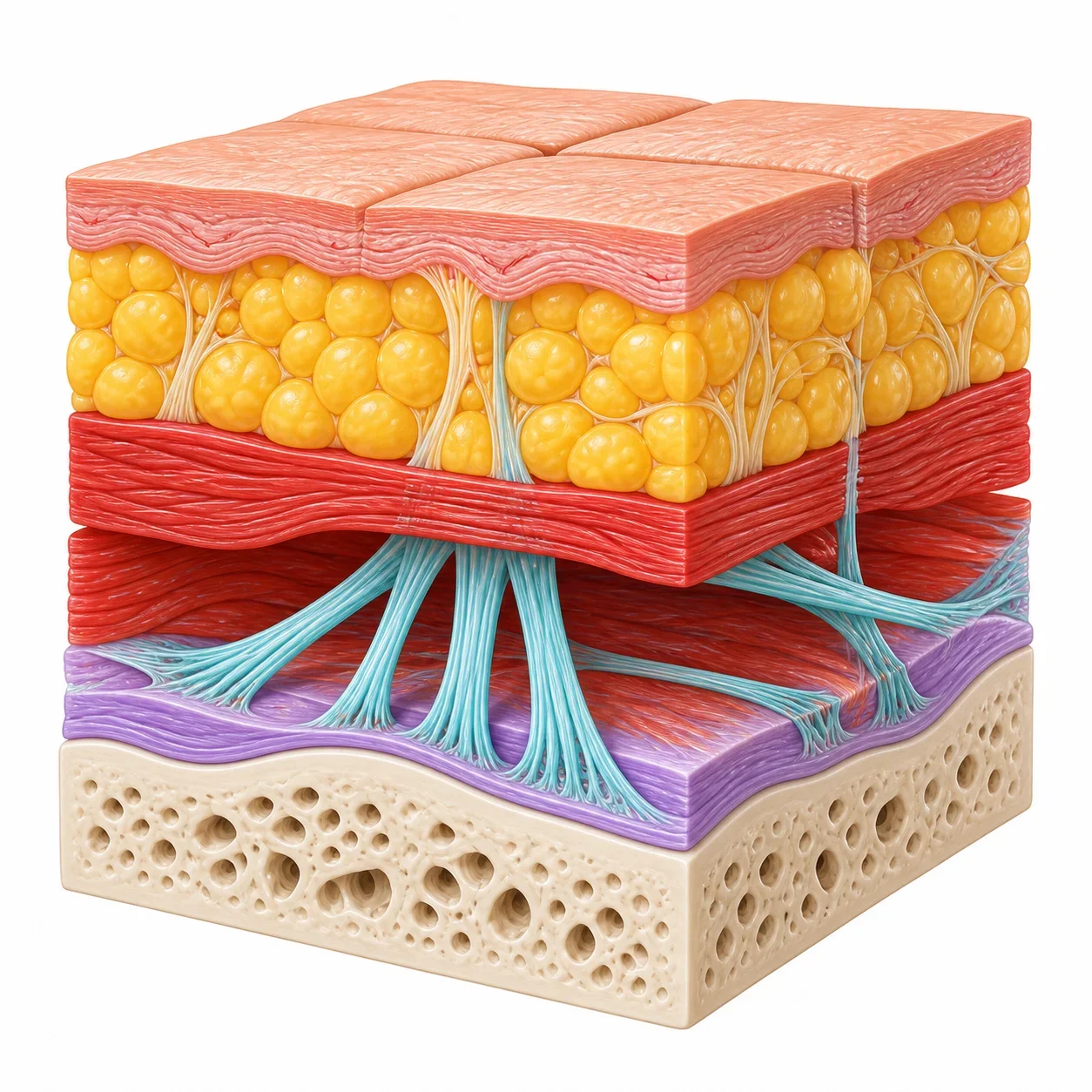
The face sags with age not simply because the skin stretches. Beneath the skin lie the fat layer, the SMAS (superficial musculoaponeurotic system), the facial expression muscles, and the retaining ligaments that hold this soft tissue to the bone and deep fascia.

In youth, these structures hold the facial tissue stably in a high position. Over time, however, the skin loses elasticity, the fat and SMAS layer shift downward, and the tissue sags and folds around the retaining ligaments, producing nasolabial folds, deep cheek sagging, marionette lines, and jowls.

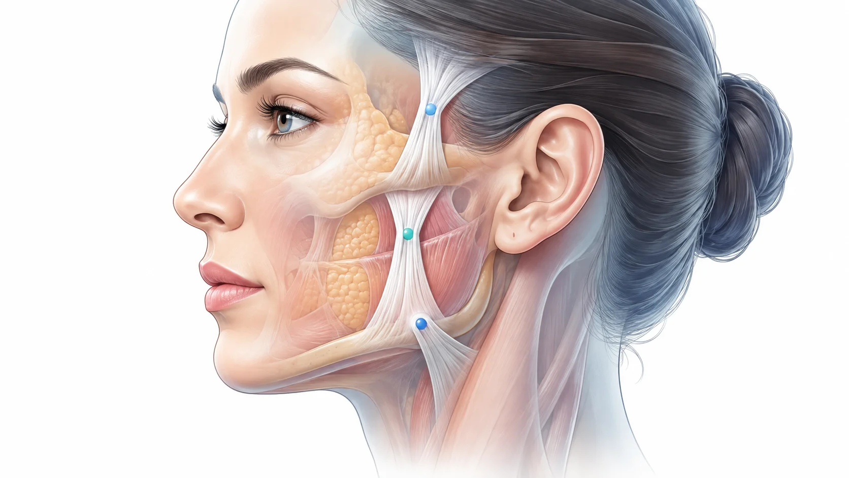
The retaining ligaments of the face are important anchoring structures that limit sagging by connecting and fixing soft tissue, including the skin, subcutaneous fat layer, SMAS, and facial expression muscles, to the facial bones.

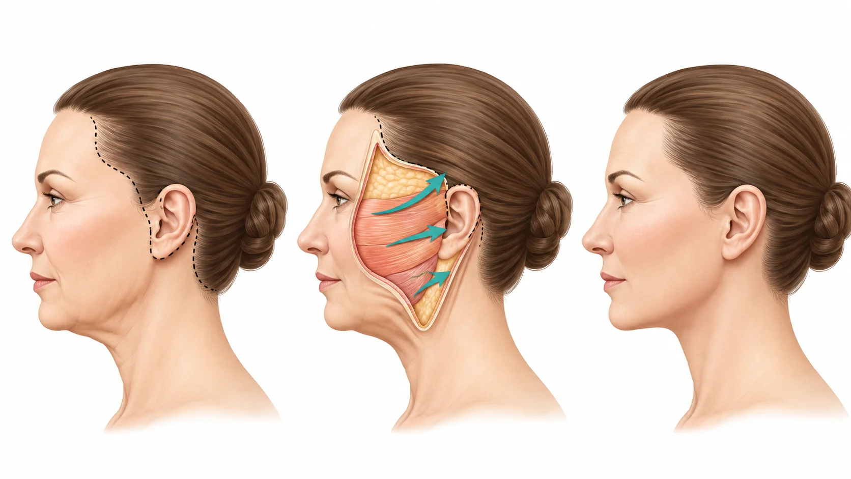
A deep plane facelift can therefore be described as a surgery that does not simply pull the skin backward. Instead, it enters the layer deeper than the SMAS, releases the retaining ligaments that restrict the movement of the sagging tissue to the extent needed, and then repositions the skin, fat, and SMAS as a single composite unit to their original, youthful position.

* A deep plane facelift is a surgery that moves the deep layers of the face together, seeking a more natural, structural age-reversing effect.

What Is a Deep Plane Facelift?

Rather than pulling the skin alone, this facelift dissects and repositions the sagging retaining ligaments and SMAS layer together
to make the face look naturally more youthful.

1Why Is It Needed?

  • With age, not only the skin but also the SMAS layer,
    retaining ligaments, and fat layer sag together.
  • Nasolabial folds, deep cheek sagging, jawline sagging,
    and marionette lines may become more noticeable.
  • Pulling the skin alone can leave a strongly pulled look
    and may not last long.
  • A deep plane facelift corrects the deeper layers as well,
    aiming for natural-looking change.

2How Does It Work?

Aged Facial Layers
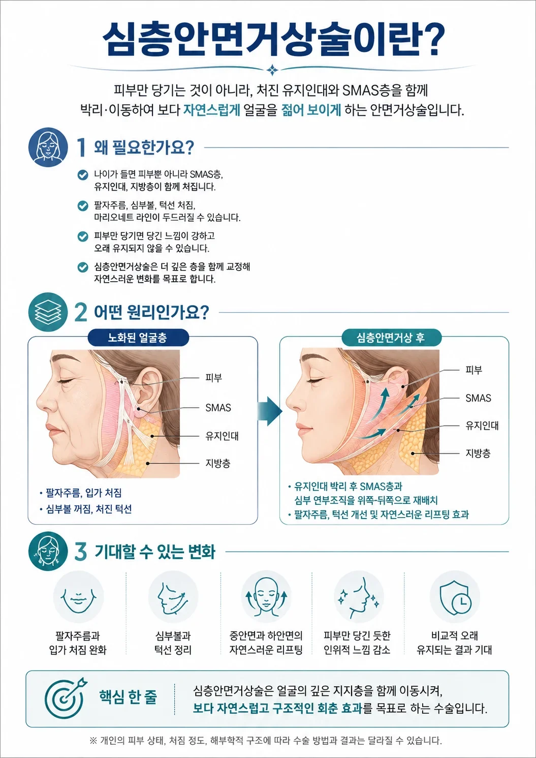
Diagram of aged facial layers
  • Skin
  • SMAS
  • Retaining Ligaments
  • Fat Layer
  • Nasolabial folds, sagging around the mouth
  • Deep cheek hollowing, sagging jawline
After a Deep Plane Facelift
Diagram of facial layers after a deep plane facelift
  • Skin
  • SMAS
  • Retaining Ligaments
  • Fat Layer
  • After dissecting the retaining ligaments, the SMAS layer and
    deep soft tissue are repositioned upward and backward
  • Improved nasolabial folds and jawline with a natural lifting effect

3Changes You Can Expect

  • Softer nasolabial folds and
    less sagging around the mouth
  • A more defined deep cheek
    and jawline
  • A natural lift of the
    midface and lower face
  • Less of the artificial look
    of skin-only pulling
  • Results expected to last
    a relatively long time

※ Surgical methods and results may vary depending on individual skin condition, degree of sagging, and anatomical structure.
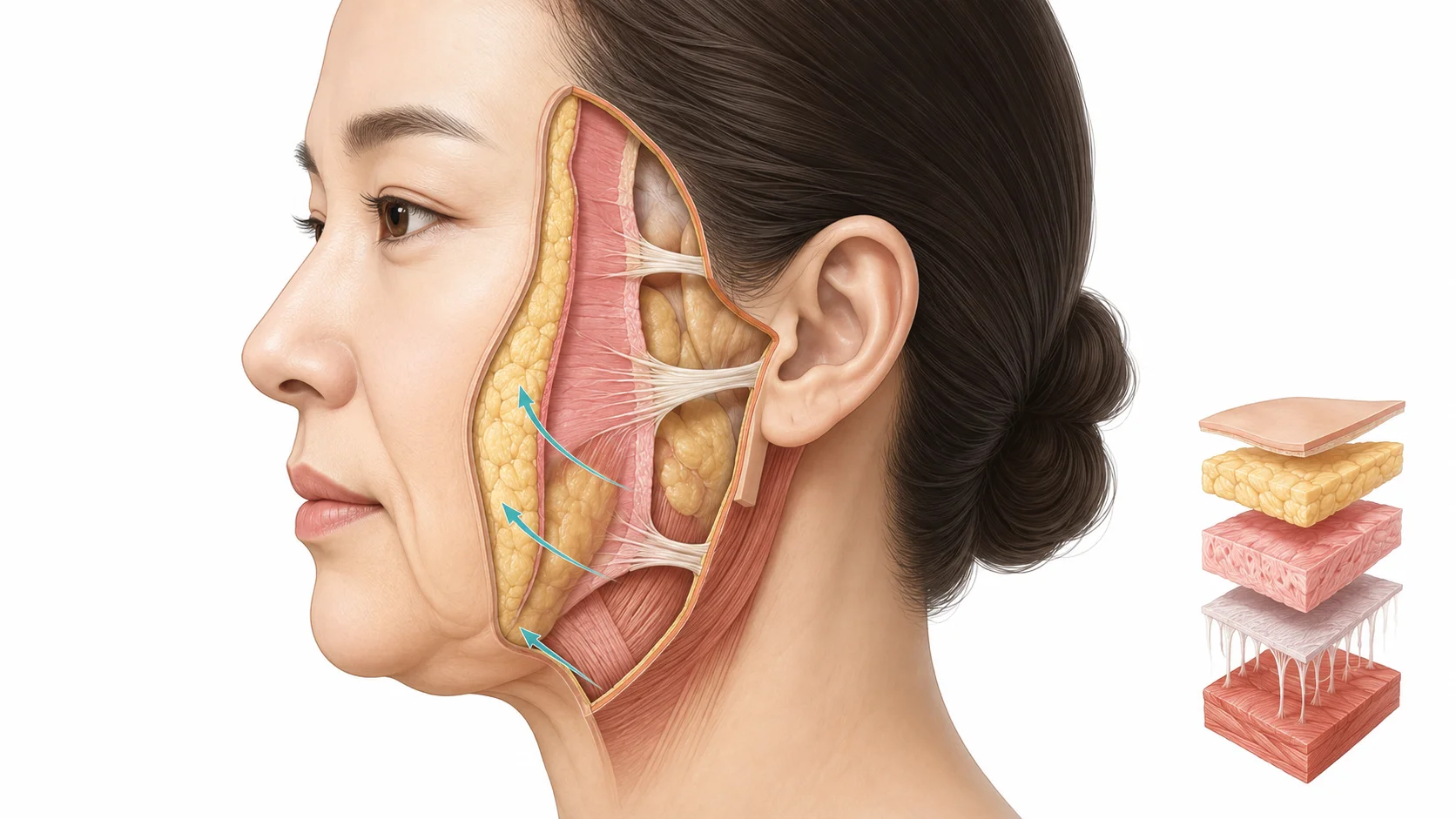
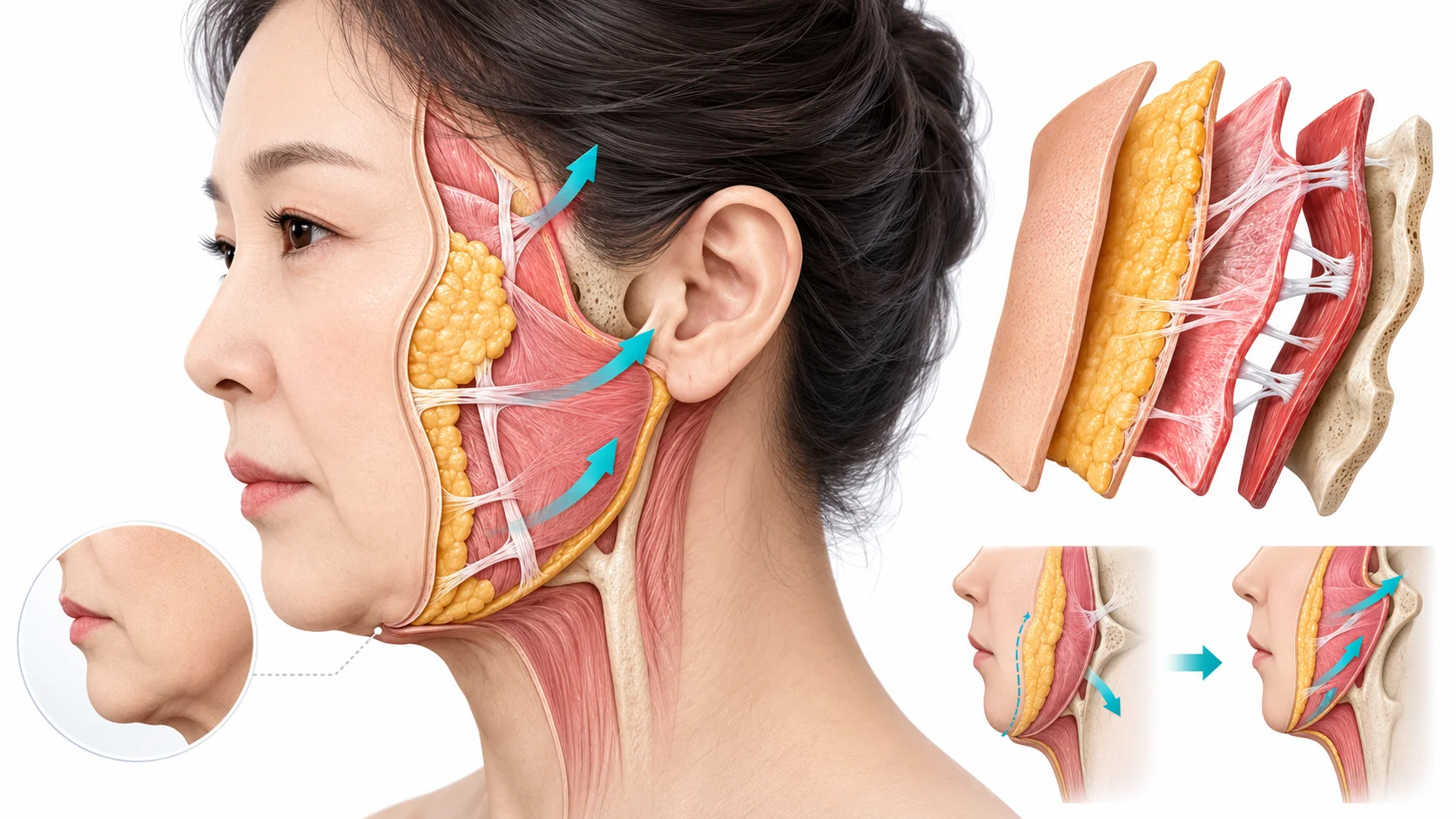
1. Facial Sagging Is Not Just a “Skin Problem”

As the fat, SMAS, and retaining ligaments beneath the skin descend together, the facial contour breaks down.

1. Facial Sagging Is Not Just a “Skin Problem”

Changes Caused by Aging

As midface fat and the SMAS shift downward, nasolabial folds, deep cheek sagging, sagging around the mouth, and jowls along the jawline appear.

Role of Retaining Ligaments

Retaining ligaments are anchoring structures that hold the facial soft tissue to the bone and deep fascia. They limit the movement of sagging tissue and can form the boundaries of wrinkles and contour irregularities.

The Goal of a Deep Plane Facelift

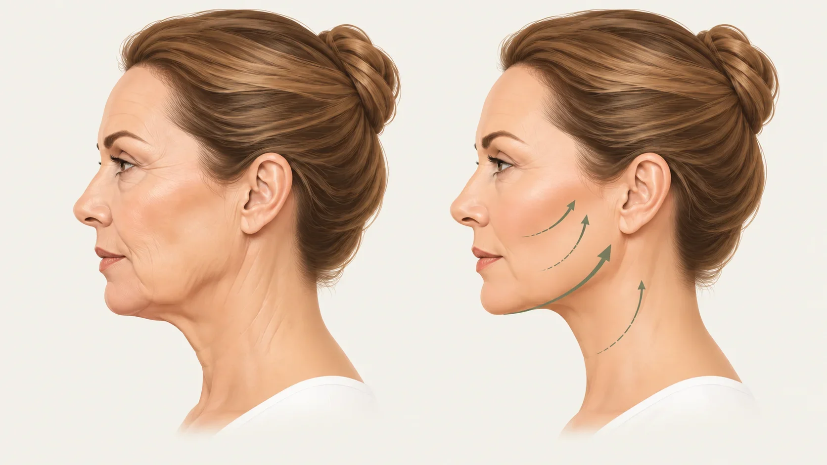
By releasing the retaining ligaments in the deep dissection plane, the tissue is not “pulled by force” but made mobile and then repositioned in a more natural direction (upward and outward: the superolateral direction).

2

2. Core Principles of the Deep Plane Facelift

In the appropriate plane beneath the SMAS, the retaining ligaments are released and the skin-fat-SMAS composite is moved together.

Not a “surgery that pulls hard,” but a “surgery that releases the tethering structures and lifts”

  • Lifting force is transmitted to the deep supporting layers, not placed as tension on the skin alone
  • By lifting the midface, lower face, and neck as a single flap, it aims for continuous contour improvement
  • Releasing the retaining ligaments increases tissue mobility, allowing natural rearrangement of the SMAS layer and the subcutaneous tissue layer
  • The skin is tailored last, without excessive tension
2. Core Principles of the Deep Plane Facelift

How It Differs from a Traditional Facelift

In a traditional skin-focused facelift, the skin is dissected relatively widely, then pulled toward the ear, and the excess skin is removed.

However, the deeper SMAS and fat tissue remain where they were, and heavy tension falls on the skin alone. As a result, the face may look pulled sideways, the skin may dimple where the retaining ligaments tether it and create contour irregularities, and tension in the skin around the ear may cause a pixie ear deformity, in which the earlobe is pulled downward.

A deep plane facelift, by contrast, is characterized by moving the deep soft tissue, including the SMAS and fat, as a single unit rather than pulling hard on the skin alone.

If a skin-only facelift is a “surgery that pulls the surface skin by force,”

a deep plane facelift is a “surgery that gently repositions the deep soft tissue of a sagging face.”

3

3. Locations and Names of the Retaining Ligaments

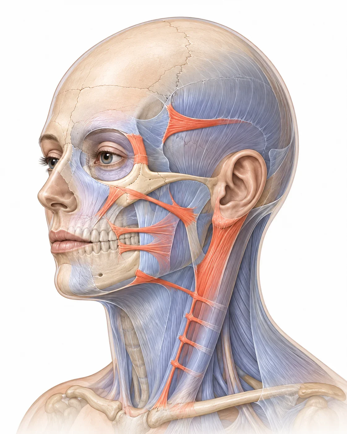
The main ligaments form a continuous series of fixation points from the temporal region to the neck.

The main ligaments form a continuous series of fixation points from the temporal region to the neck.
  1. 1

    Temporal Adhesion / Septum

    TLA · STS · ITS

  2. 2

    Periorbital Fixation

    TTL · ORL · LOT (Orbital retaining ligament)

  3. 3

    Zygomatic Complex

    Zygomatic ligaments

  4. 4

    Maxillary / Masseteric Complex

    Maxillary · Masseteric

  5. 5

    Mandibular Attachment

    Mandibular attachment

  6. 6

    Parotid / Auricular Fixation

    PAF · PAL · Lore’s/TPF

  7. 7

    Posterolateral Cervical Adhesion

    CRL / Posterior adhesion

  8. 8

    Proposed Structures

    HPL · CMAS: Defined Differently Across the Literature

AI-generated educational location diagram · The actual number and shape of bundles vary by individual

Educational anatomy material · In actual surgery, individual variation and direct vision take priority

04

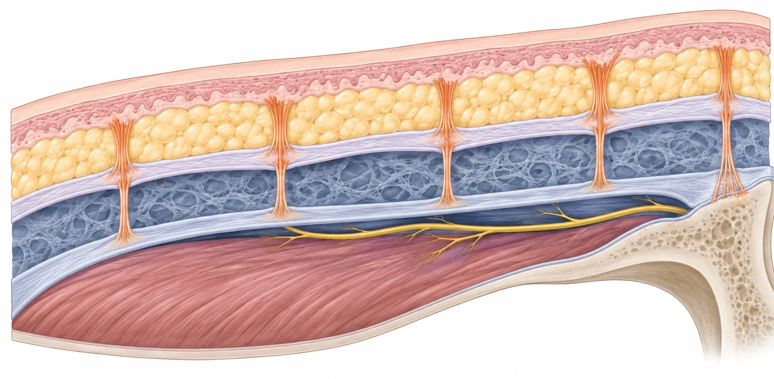
Retaining Ligaments Are Not “Strings” but a Support Network Linking the Layers

Retaining Ligaments Are Not “Strings” but a Support Network Linking the Layers

AI-generated educational cross-section diagram

Skin / Subcutaneous Tissue

SMAS / Platysma

Deep-plane space

Deep Fascia / Periosteum

3 Anatomical Roles

Support: maintains the position of the soft tissue

Compartment: forms the boundaries of the mobile spaces

Landmark: signals where nerves and vessels change layers

Key: read the ligaments, spaces, and structures at risk as a single 3D map.

Educational anatomy material · In actual surgery, individual variation and direct vision take priority

02

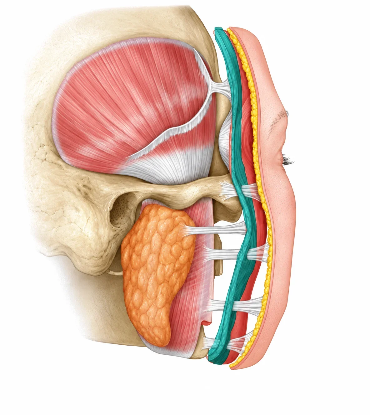
4. Anatomical Relationship Between the Retaining Ligaments and the SMAS

The retaining ligaments originate from the bone or the deepest muscle layer and run vertically through the SMAS layer up to the skin layer.

Educational diagram showing the anatomical relationship between the retaining ligaments and the SMAS layer 123 456 789 1011
  1. 1

    Skin

  2. 2

    Galea plane

  3. 3

    Sub-galea plane

  4. 4

    Zygomatic ligament
    true ligament

  5. 5

    Masseteric ligament

  6. 6

    SMAS plane

  7. 7

    Sub-SMAS plane

  8. 8

    Mandibular ligament
    true ligament

  9. 9

    Deep fascia plane

  10. 10

    Parotid gland

  11. 11

    Masseter

(1) Upper Face Retaining Ligaments: Defining the Boundaries of Forehead, Temple, and Lower Eyelid Dissection

StructureMain LocationSurgical Significance
Temporal ligamentous adhesionNear the superior temporal lineStrong fixation point of the forehead-temple flap
Superior temporal septumAround the superior temporal lineBoundary between the frontal and temporal dissection spaces
Inferior temporal septumAbove the zygomatic archInferior boundary close to the frontal branch of the facial nerve
Lateral orbital thickeningLateral orbital rim periosteumFixes the lateral brow and orbicularis oculi
Orbicularis retaining ligamentOrbital rim periosteumUpper boundary of the lower eyelid and the prezygomatic space

Names and extents differ across the literature, so the “layer and fixation site” should be confirmed together.

Retaining Ligaments of the Face and Neck · 4

(2) Retaining Ligaments of the Midface and Lower Face

Retaining ligaments restrict tissue movement, but they are also important surgical landmarks close to the facial nerve.

1. Zygomatic Ligament

Anchors the cheekbone and midface tissue. If the release is insufficient, the sagging tissue above the nasolabial fold may not move sufficiently.

2. Masseteric Ligament

Involved in the boundary between the cheek and the lower face. Because facial nerve branches lie nearby, it must be dissected carefully under direct vision in the correct plane.

3. Mandibular Ligament

Involved in the jawline and jowl formation. It is approached to the extent needed to smooth the lower face contour.

(2) Retaining Ligaments of the Midface and Lower Face

Safety Points

Retaining ligament release is the step that gains mobility, and also a highly demanding step that requires consideration of the course of the nerves.

8

- Midface mobility depends on releasing the zygomatic and masseteric ligaments

Zygomatic Retaining Ligaments

  • Anchor the SMAS at the zygoma and anterior to the zygomatic arch
  • Inferior boundary of the prezygomatic space
  • Key to moving the midface and malar fat pad

Masseteric Retaining Ligaments

  • Run from the fascia at the anterior border of the masseter to the superficial layer
  • Observed as upper and lower series
  • Closely related to facial nerve branches

Principles of a Safe Approach

  1. ①

    Open the space first

    Pre-zygomatic / Pre-masseter space

  2. ②

    Identify the boundary ligaments

    Expose the fiber bundles under direct vision

  3. ③

    Anticipate the course of the nerves

    Protect the branches deep to the ligaments

  4. ④

    Release only as much as needed

    Secure the mobility needed for the target vector

Retaining Ligaments of the Face and Neck · 5

- The lower face is determined by the release of the mandibular ligament.

Fixation Point

Mandibular Retaining Ligament

Runs from the periosteum of the anterior mandible to the superficial layer, holding the jawline in a constant position.

Aging Changes

  • Descent of the soft tissue in front of and behind the ligament
  • The fixation point stays relatively in place, accentuating the boundary
  • Jowls and the pre-jowl depression appear together

Surgical Significance

More than simple skin traction, sufficient mobility of the SMAS-platysma complex and an anatomical understanding of the area around the mandibular septum are important for improving the jawline.

Retaining Ligaments of the Face and Neck · 6

(3) Retaining Ligaments of the Neck: Structures That Anchor the Platysma to the Deep Cervical Fascia

Platysma–auricular ligament

Posterior anchoring structures below the ear and around the parotid gland and mastoid process. Their names partly overlap with Lore’s fascia and others.

Cervical retaining ligaments

Connects the lateral platysma to the deep cervical fascia over the SCM, limiting superior and posterior redraping.

Purpose of the Release

To secure mobility of the platysma-SMAS complex and improve the neck contour and banding

(3) Retaining Ligaments of the Neck: Structures That Anchor the Platysma to the Deep Cervical Fascia

Retaining Ligaments of the Face and Neck · 7

5. Deep Plane Facelift: Releasing the Retaining Ligaments

Layer Structure and Roles

  • Skin
  • Subcutaneous fat
  • SMAS (fascial layer)
  • Retaining ligament
  • Periosteum
Diagram of the layered structure of skin, fat, SMAS, retaining ligaments, and periosteum
Educational facial diagram comparing no retaining ligament release with a deep plane lift

Without Retaining Ligament Release
(Skin Pull Only)

Pulling only the skin and subcutaneous fat
is blocked by the retaining ligaments,
giving a less natural result

After Retaining Ligament Release
(Deep Plane Lift)

Releasing the retaining ligaments and lifting
the SMAS from the deep layer
gives a natural, long-lasting result

AI-generated comparison image: before
Before (Concept Image)
AI-generated comparison image: after
After (Concept Image)

These are AI-generated comparison images, not actual before-and-after photos of a patient.

6. Surgical Process

The order is: consultation → incision → deep plane dissection → retaining ligament release → lift and fixation → skin tailoring.

6. Surgical Process

01 Consultation & Design

We assess the location of sagging, skin thickness, fat volume, the condition of the neck and jawline, and previous surgeries.

02 Incision Planning

Incisions are planned along the hairline and the creases around the ear so that scars are concealed.

03 Deep Plane Dissection

We enter the appropriate plane beneath the SMAS so that the sagging tissue can be moved sufficiently.

04 Retaining Ligament Release

The anchoring structures in the zygomatic, masseteric, and mandibular regions are carefully dissected to the extent needed.

05 Lift & Fixation

The SMAS/platysma and soft tissue are repositioned upward and backward and secured.

06 Skin Tailoring

Excess skin is trimmed naturally and sutured without tension.

The actual extent of surgery varies with each person's pattern of aging and degree of neck sagging.

13

7. Wrinkles Improved by a Deep Plane Facelift

It is effective for structural sagging of the midface and lower face, but it is not a surgery that completely removes every wrinkle.

Educational comparison diagram showing facial sagging and the direction of lift

Nasolabial Folds

Softened by reducing the weight of the sagging cheek tissue. Deep skin creases may remain.

Deep Cheek

Cheek fat and SMAS that have descended are repositioned to restore a smooth curve.

Jawline & Jowls

Improving the sagging around the mandibular retaining ligament can give a more defined jawline.

Marionette Lines

The heavy look below the corners of the mouth is reduced, but deep wrinkles do not disappear completely.

Neckline

When the platysma and neck sagging are also involved, a neck lift/platysmaplasty may be needed as well.

Skin Texture & Fine Lines

Lifting surgery alone does not resolve all pores, pigmentation, and fine lines.

Please Note

It works “in the direction of returning to a younger face,” but it is not a “surgery that erases every wrinkle.” For fine lines caused by skin aging in particular, the effect may be amplified by targeting the skin layer with treatments such as skin boosters, Er:YAG laser, Ultherapy, and XERF rather than with a lift.

14

8. Advantages · Disadvantages · Recovery Process

Explaining the advantages and limitations in a balanced way helps set the patient's expectations realistically.

Advantages

  • Improves the midface and lower face together
  • Relatively less of a skin-only pulled look
  • Helps reduce skin tension around the ear
  • Long-term improvement can be expected in suitable patients
  • - Maintains a thicker, more physiologic SMAS-platysma flap
  • - Maintains midface volume with natural boundary lines
  • - Longer-lasting results from a lift that is more vertical than horizontal
  • - Less unnatural pulling by minimizing lateral traction

Drawbacks & Limitations

  • Deep surgical field and high anatomical difficulty
  • Possible hematoma, infection, scarring, and asymmetry
  • Possible reduced sensation or temporary weakness of facial expression
  • Fine lines, skin texture, and pigmentation require separate treatment
  • Aging continues after surgery

Recovery

  • First 1-2 weeks: swelling, bruising, tightness
  • Several weeks: major swelling subsides, minor swelling persists
  • Several months: firmness and sensory changes gradually improve
  • Sudden pain or one-sided swelling: seek care immediately
  • Follow your medical team's guidance on when to wash your face, exercise, and dye your hair

15

9. Key Points of the Deep Plane Facelift

More important than the name Deep Plane are the correct dissection plane, safe anatomy, and individualized vector design.

A deep plane facelift is not a surgery that pulls the skin tight; it repositions the deep soft tissue of the face, which has descended with aging, closer to its original anatomical position.

9. Key Points of the Deep Plane Facelift

Suitable Patients

Those whose whole face has sagged together, with nasolabial folds, deep cheek sagging, jowls, and neck sagging, and who want natural structural restoration rather than a skin-only pulled look

Individualized Planning

The method varies with skin thickness, fat volume, bone structure, retaining ligament strength, neck condition, and any previous surgery.

Essence of the Surgery

The key is retaining ligament release + SMAS/soft tissue repositioning + tension-free skin tailoring.

Realistic Expectations

Rather than removing every wrinkle, including fine lines, the goal is to return the direction of facial sagging and the contour to a more youthful position.

Conclusion: “We aim for natural rejuvenation by repositioning the deep layers more precisely.”

16

Cases.

Before

After

Before
After
Before
After
Before
After
Before
After
Before
After
Before
After

RESUM Plastic Surgery Clinic Location

#601, 6F, 842 Nonhyeon-ro, Gangnam-gu, Seoul

6F #601, 599-4 Sinsa-dong, Gangnam-gu, Seoul

HOURS

Weekdays
10:00 ~ 20:00
Saturday
10:00 ~ 17:00
Lunch Break
1:00 PM - 2:00 PM

(No lunch break on weekends and public holidays) *Dr. Won-Suk Kook is not available on the 1st and 3rd Saturdays of each month.

Parking Information Parking is available in the building. 1 hour of free parking

Subway 5-minute walk from Exit 3 of Apgujeong Station on Line 3 (right next to the 14-story CGV building at Exit 3)

RESUM Plastic Surgery Clinic CEO Won-Suk Kook Business No. 733-11-02929 Phone 02-3445-7770