
Removal of an Extruded, Infected Implant: Case II
Removal of an Extruded, Infected Implant: Case II
(Removal of infected tissue and protruded silicon implant)

If an implant is extruding or is accompanied by swelling and tenderness, infection should be suspected.
This infection thickens the capsule and scar tissue, which often becomes a cause of contracture.
In other words, if any of the above symptoms appear after rhinoplasty, it is important to consult an experienced surgeon
without fail and have the problem addressed early.
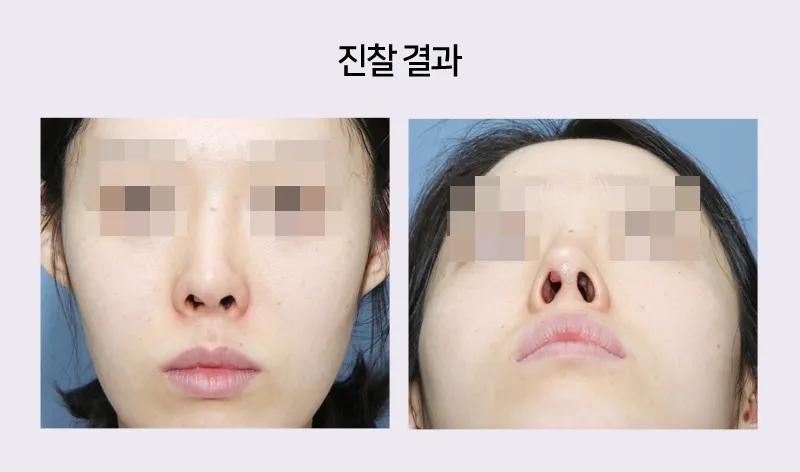
In this patient, the implant had extruded inside the right side of the nose, and a granuloma covered the area around the extruded silicone implant.
The dorsum is swollen, the bridge is thick and blunt, and the implant has shifted out of position and is crooked.
At the alae, contracture has shrunk the skin, leaving the nostrils excessively exposed.
The columella is tilted, and the two
nostrils are asymmetric as well.

The patient wanted revision surgery, but implant extrusion
means that infection has developed inside the nose, so rather than revision surgery,
the implant, capsule, scar tissue, and infected tissue must be removed first.
If, with infection present like this, revision surgery using autologous tissue or silicone
is performed, the infection may recur, so caution is required.
The surgery was to be performed through an incision inside the nose (closed approach), as follows:
1) Removal of the silicone implant
2) Removal of the capsule and scar tissue
3) Separation of the tip cartilages, followed by a derotation graft.
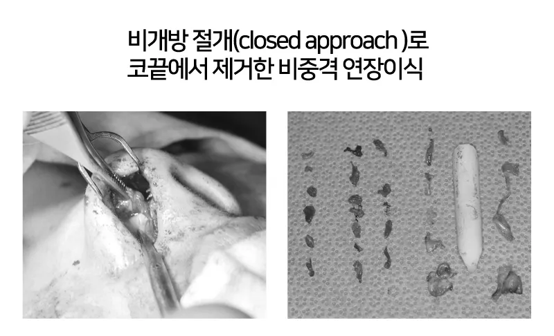
The photo on the left shows the thick granuloma and scar tissue covering the tip cartilages being removed during surgery.
The photo on the right shows the removed silicone, capsule, and scar tissue.
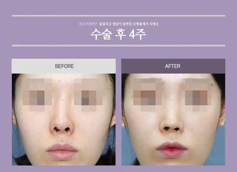
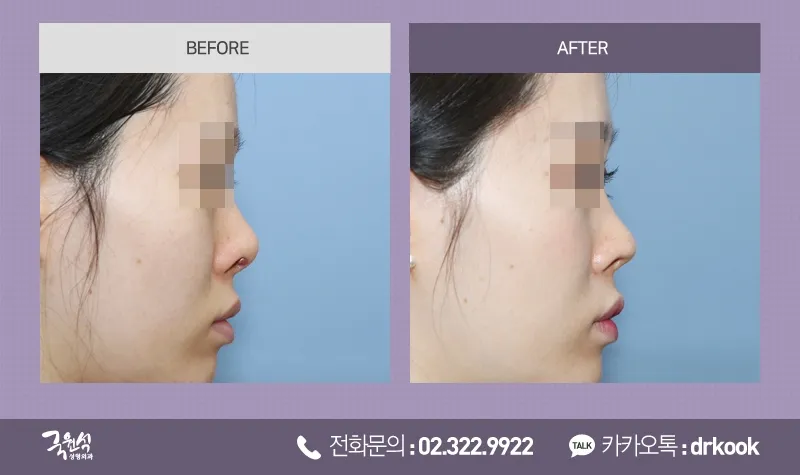
The dorsum, crooked and thick before surgery, is now slim and straight.
In addition, the alar rim had been pulled upward into a pointed shape, exposing much of the nostrils,
but after surgery the alar rim is rounded and the nostrils are covered.
The implant that had extruded inside the nose was removed cleanly, and the wound has healed well.
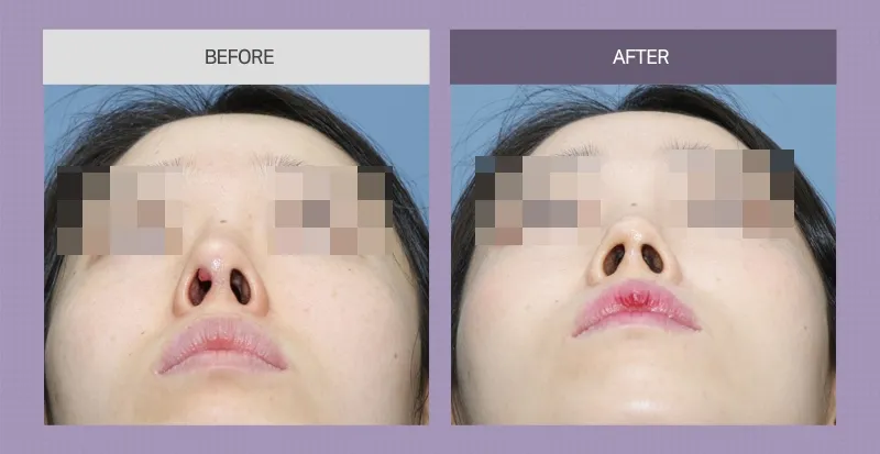
After removal of the implant that had extruded in the right nostril, the granuloma, and the scar tissue,
the overall shape of the nose, including the nostrils, bridge, and columella, was corrected,
and natural left-right symmetry was achieved.
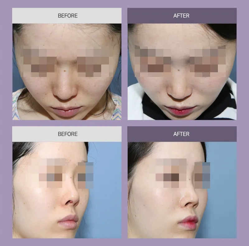
The lateral view also shows that the tip and dorsum have become slimmer.
After surgery, no contracture causing the nose to turn up occurred.
(No cephalic rotation, slimmed nasal dorsum and tip)