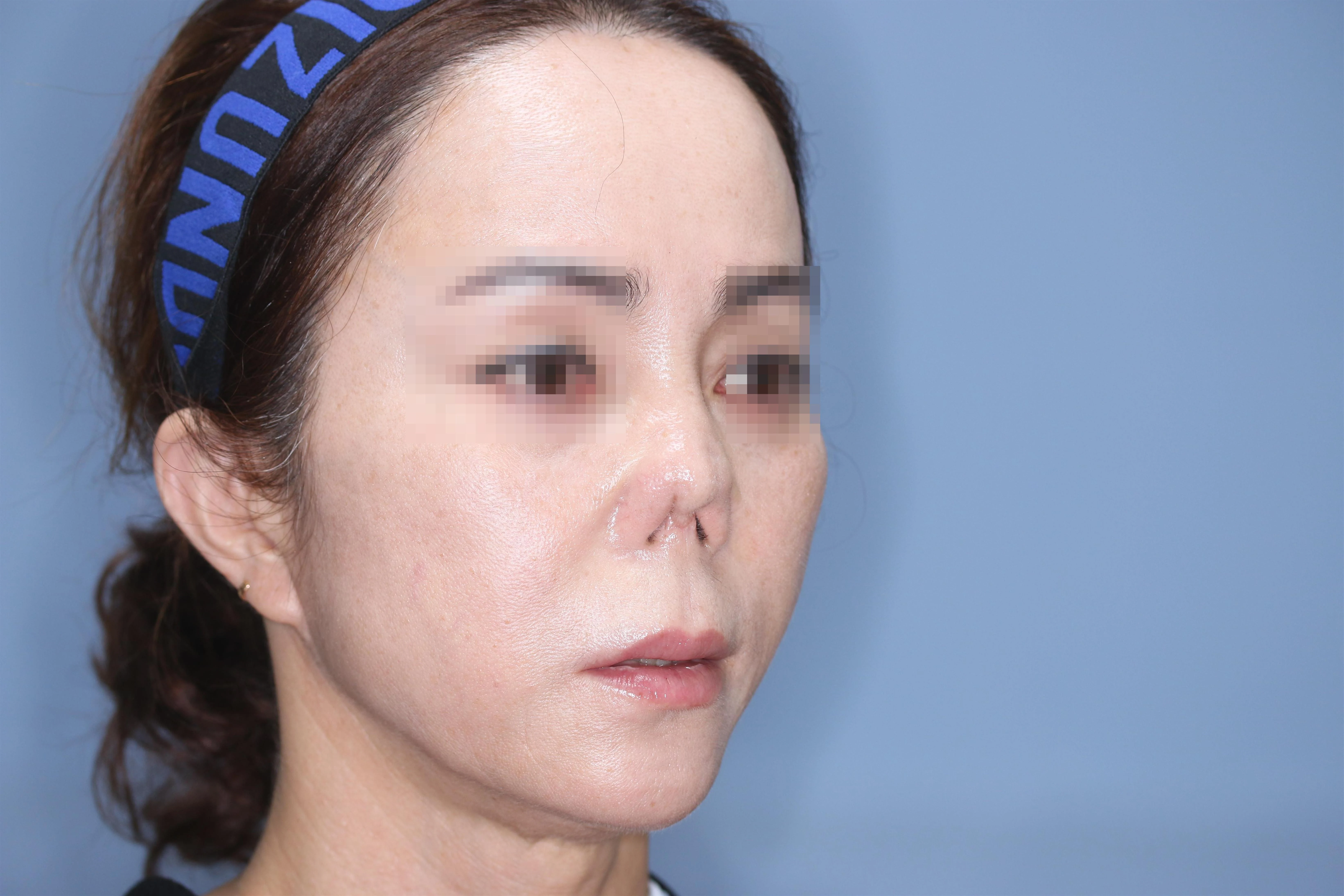
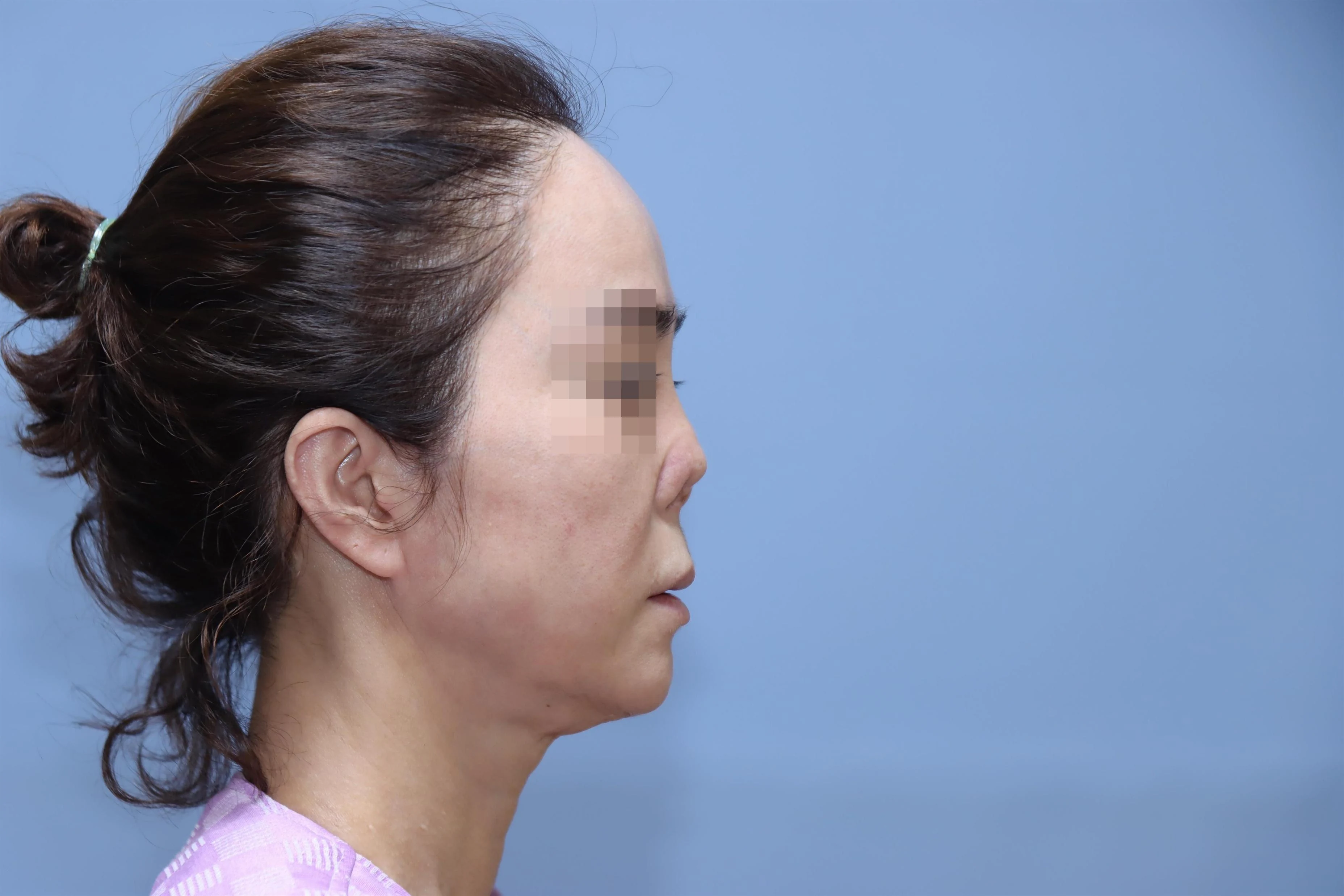
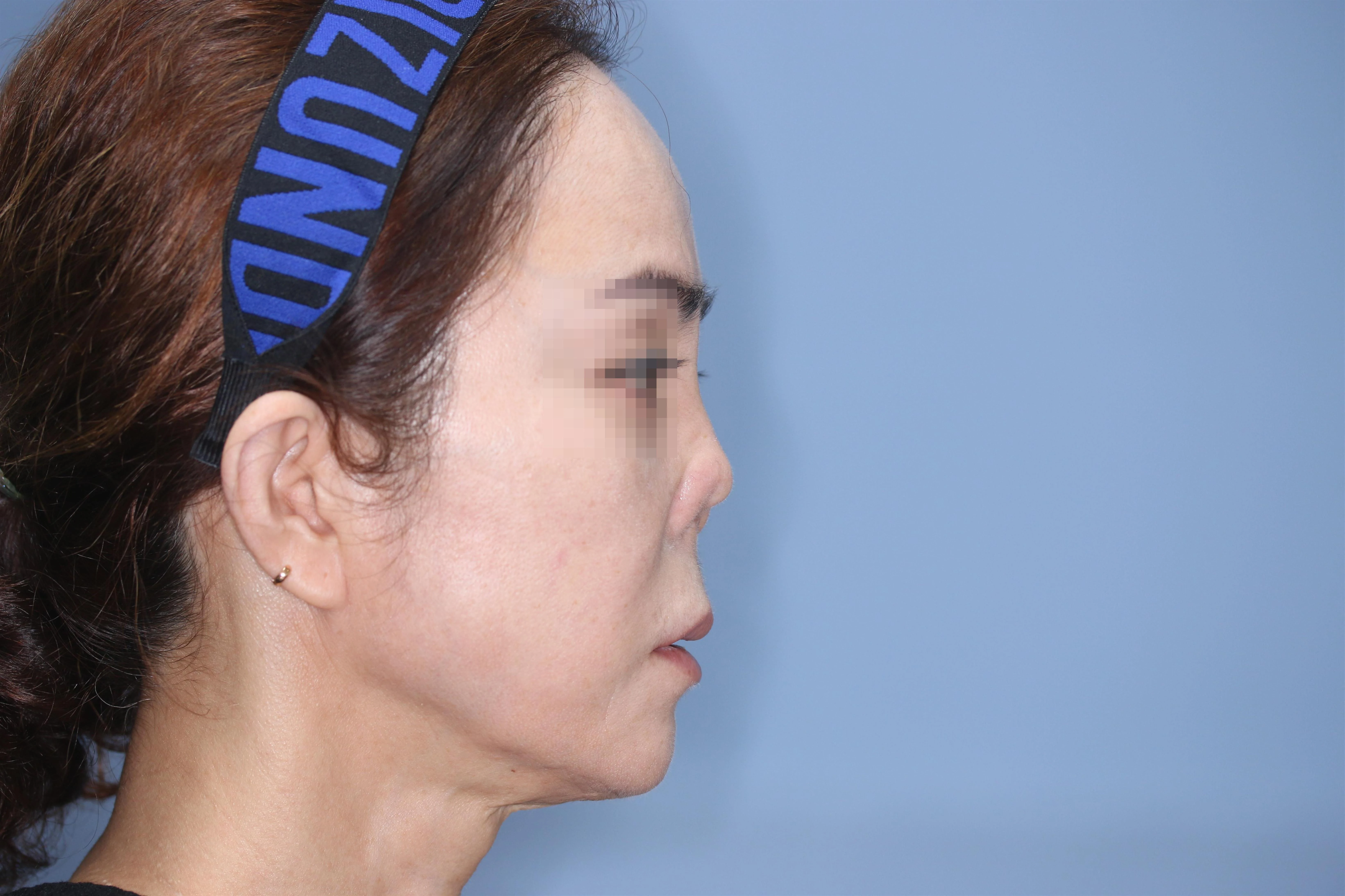
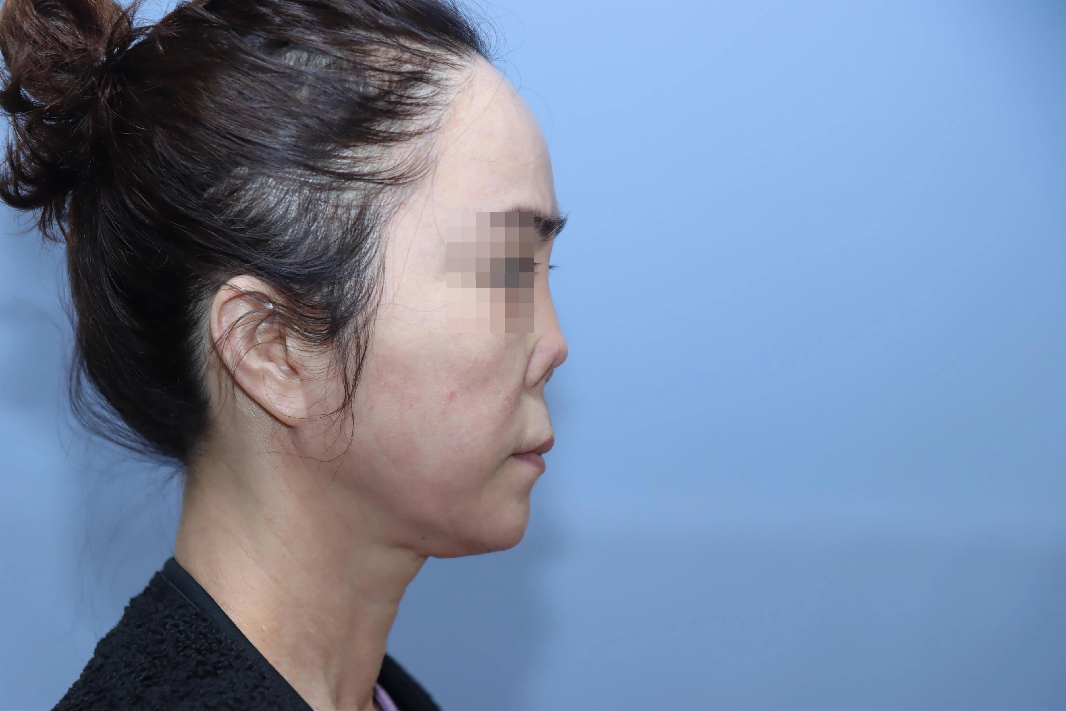
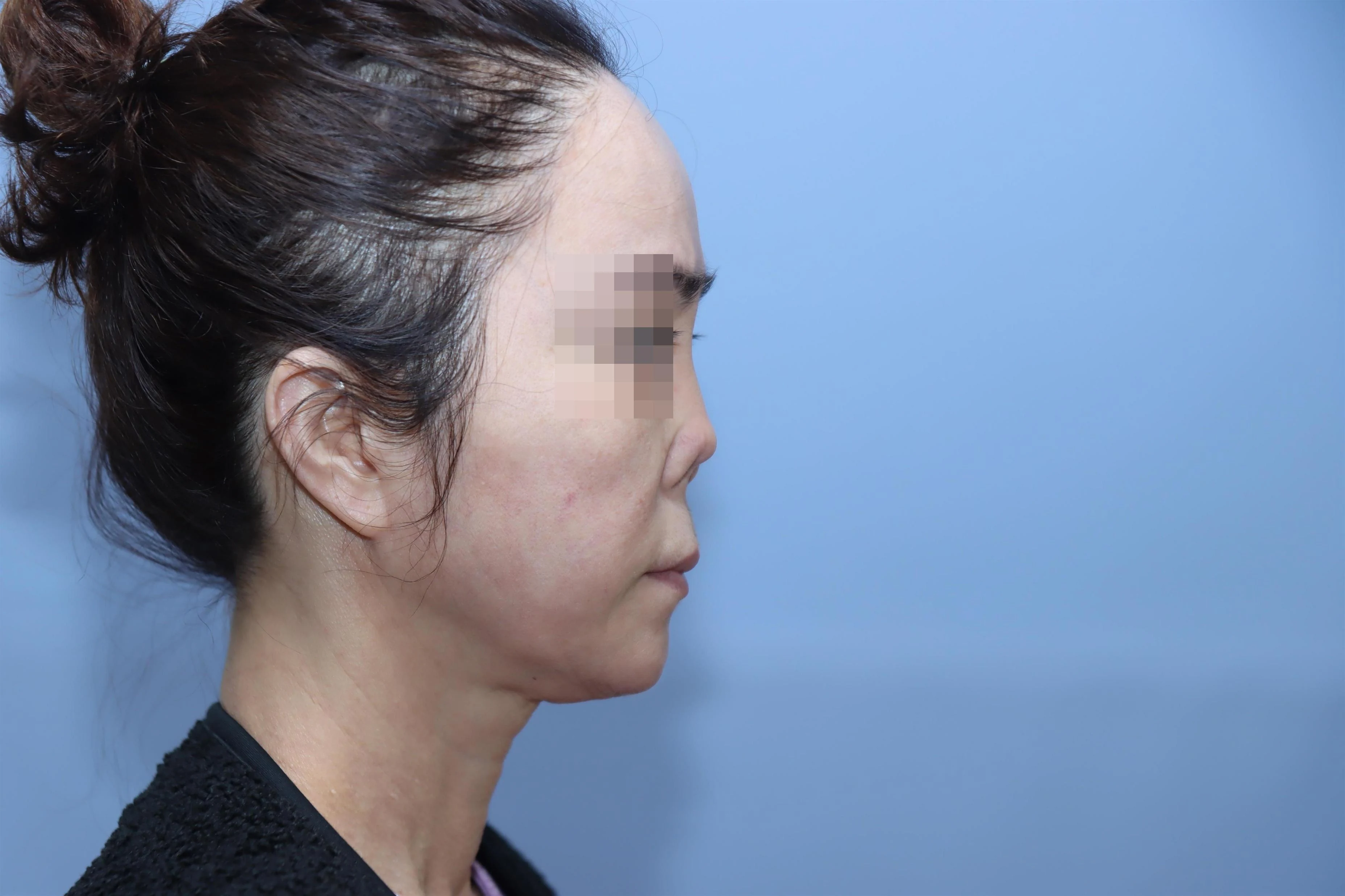
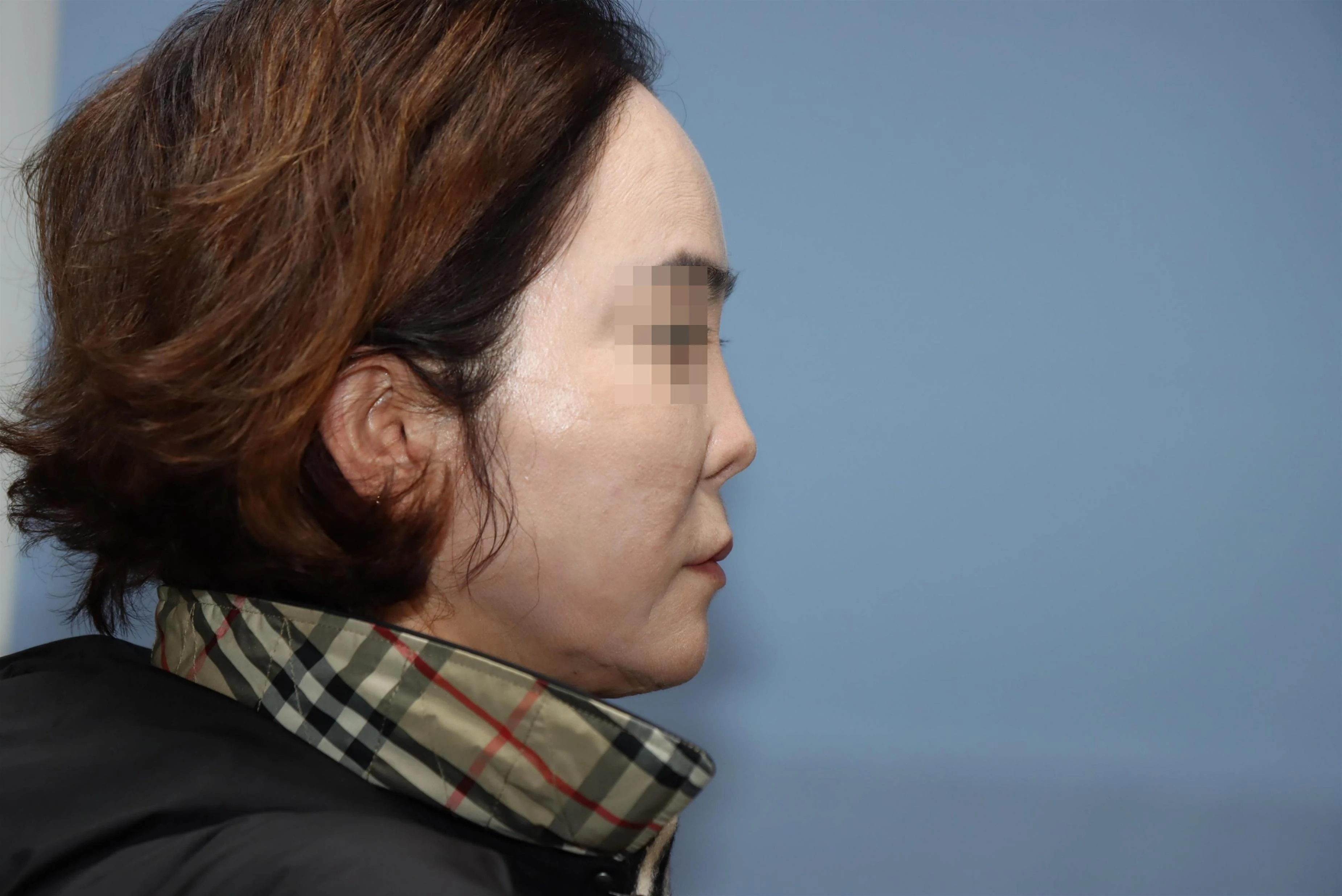
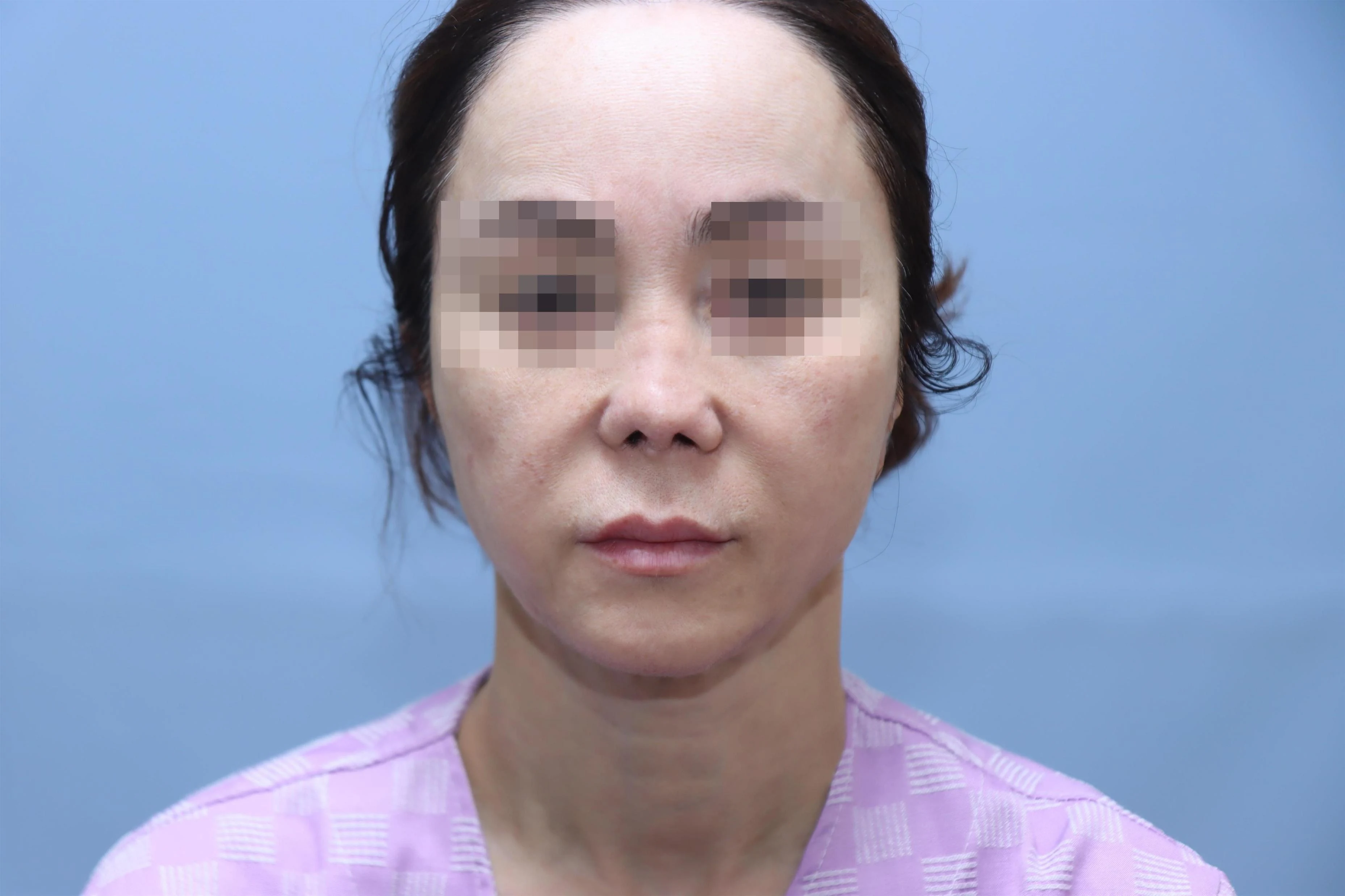
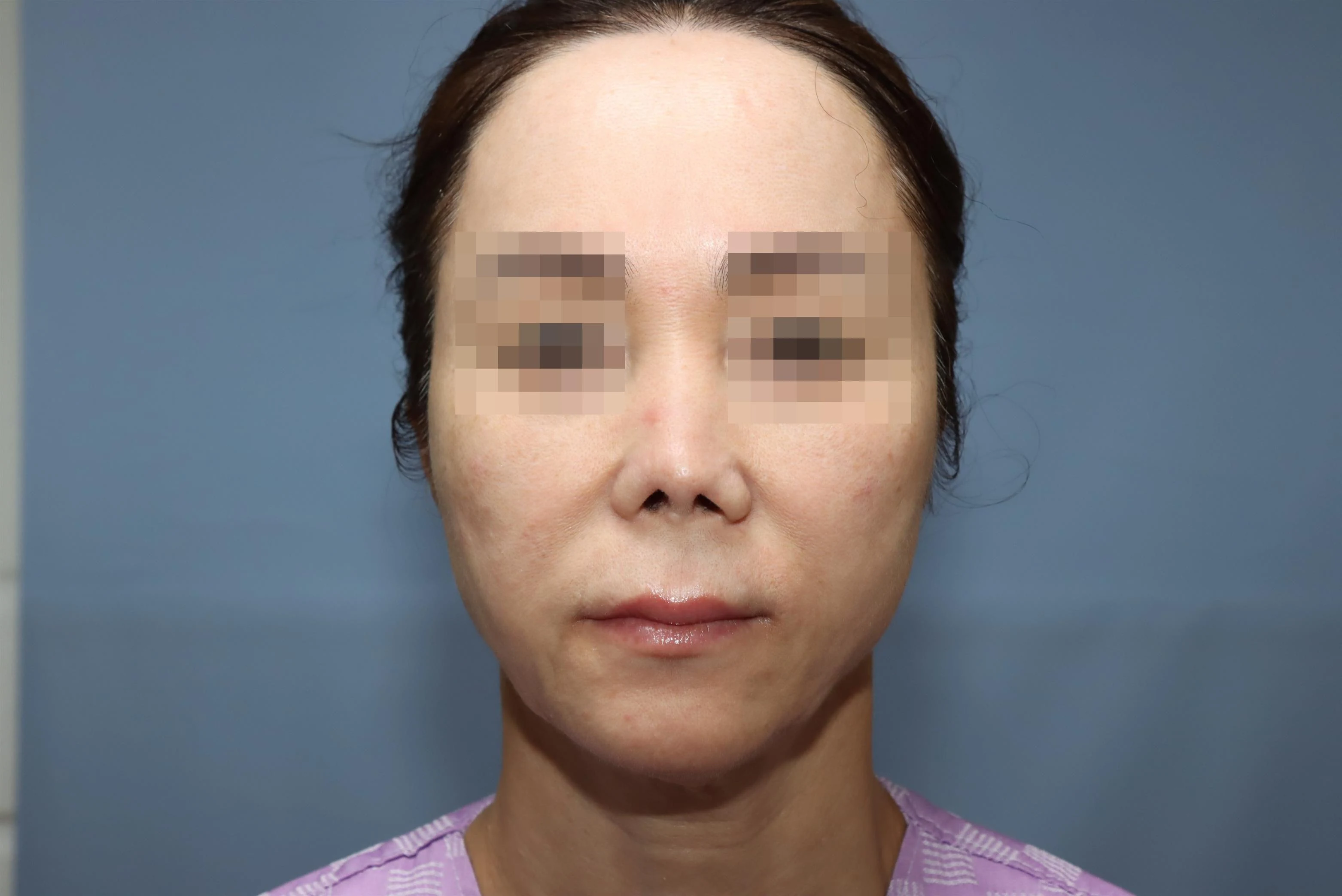
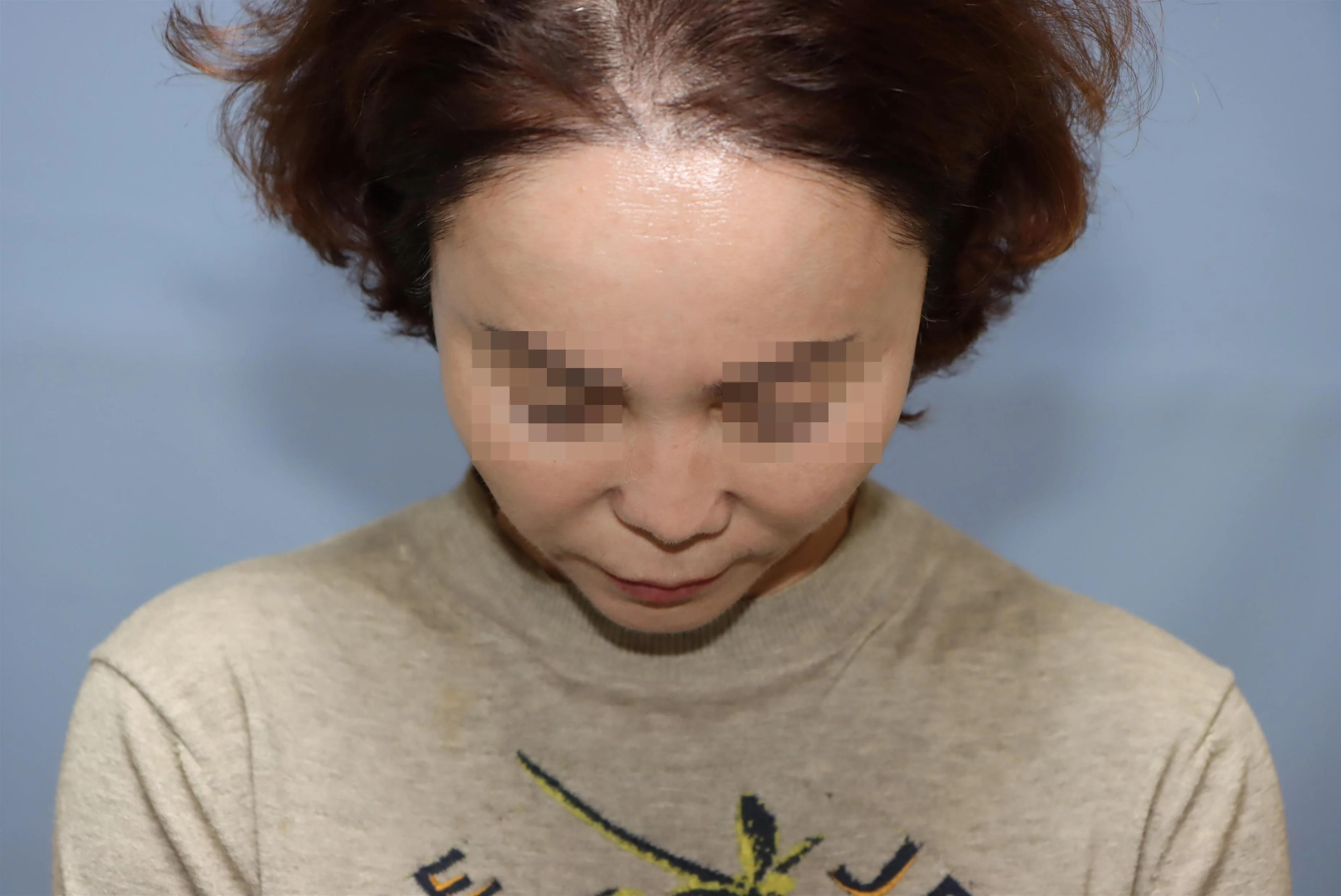
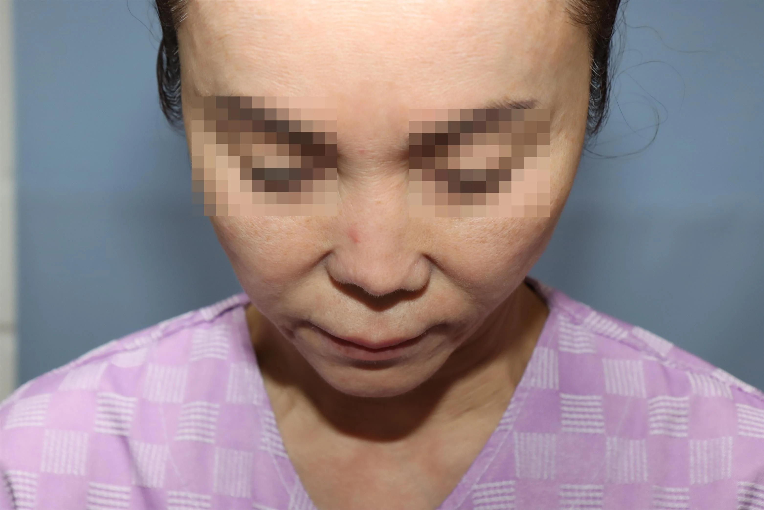
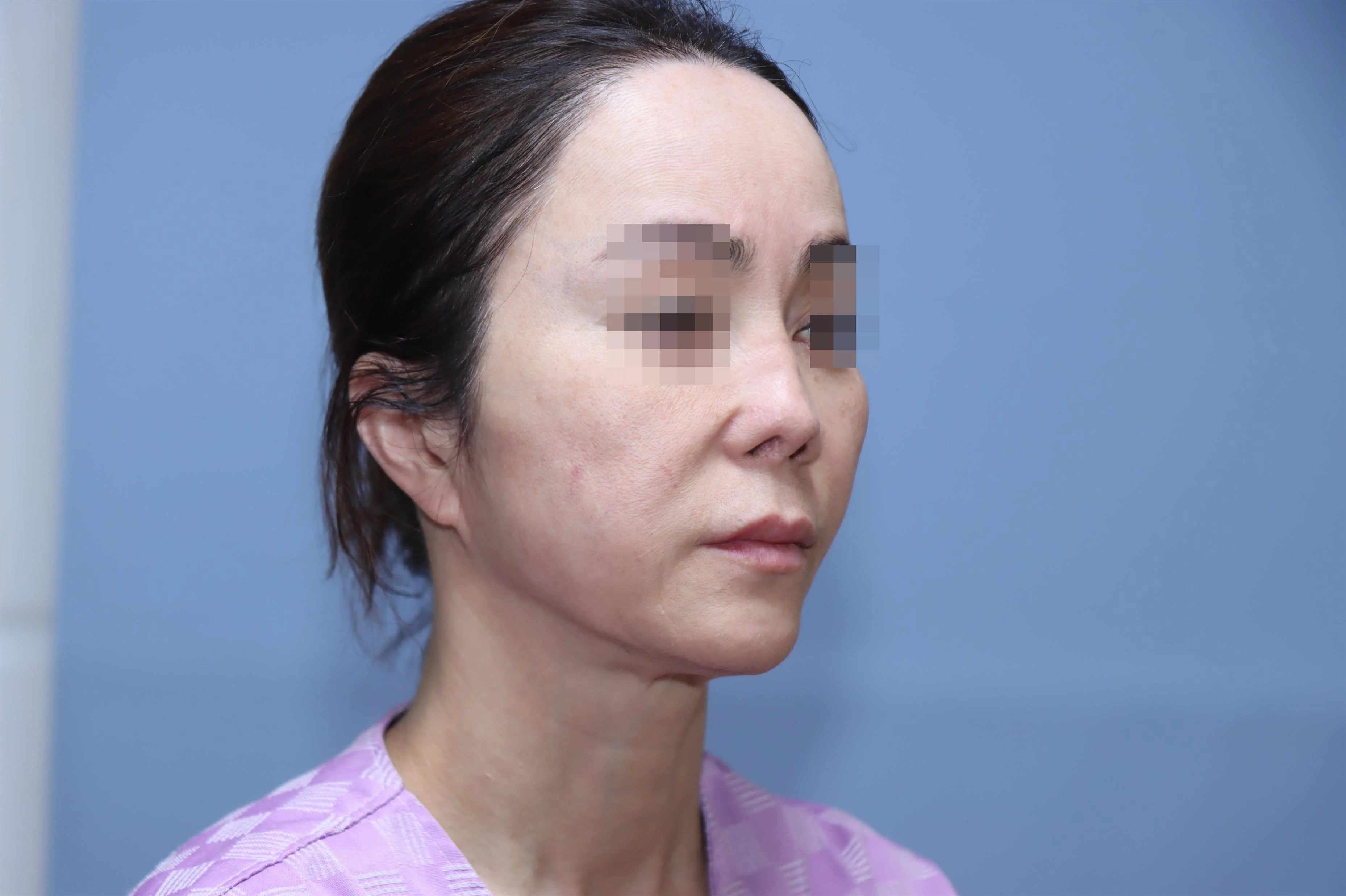
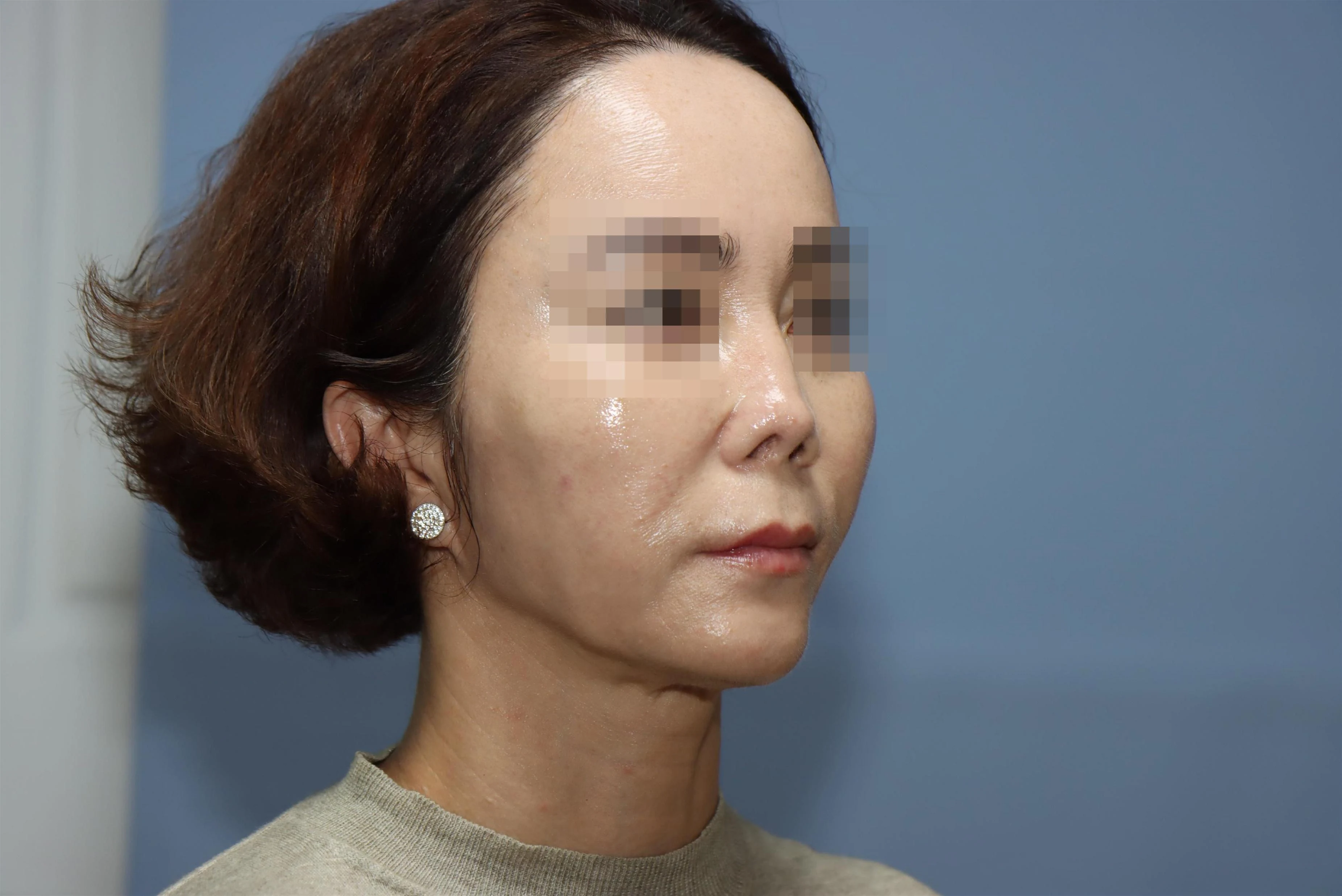
SummaryStem cell rhinoplasty is a regenerative form of rhinoplasty performed after nasal soft tissue that has become stiff and poorly supplied with blood, due to repeated surgery or implant side effects, is first restored with autologous SVF and PMedical ReviewMedical Review: Won-Suk Kook,Jin-Young Kim Last updated: 2026-09-11
SCROLL
Why Resum
OverviewBeyond the Trials and Pain Brought On by Overambition
Side effects and rhinoplasty repeated again and again because of excessive ambition and poorly performed surgery. Looking back on the course of a patient whose trials and pain finally ended after 9 surgeries, we will consider why "stem cell rhinoplasty" is needed and what it means.
As with everything, what calls for the greatest caution in rhinoplasty is overambition and formulaic surgery applied identically to everyone.
Reconstructive Surgery
* What Is Stem Cell Rhinoplasty?
SVF · PRP / PRFThe nasal skin and soft tissue damaged by repeated surgery are reinforced with stem cells, and nasal reconstruction is then completed.
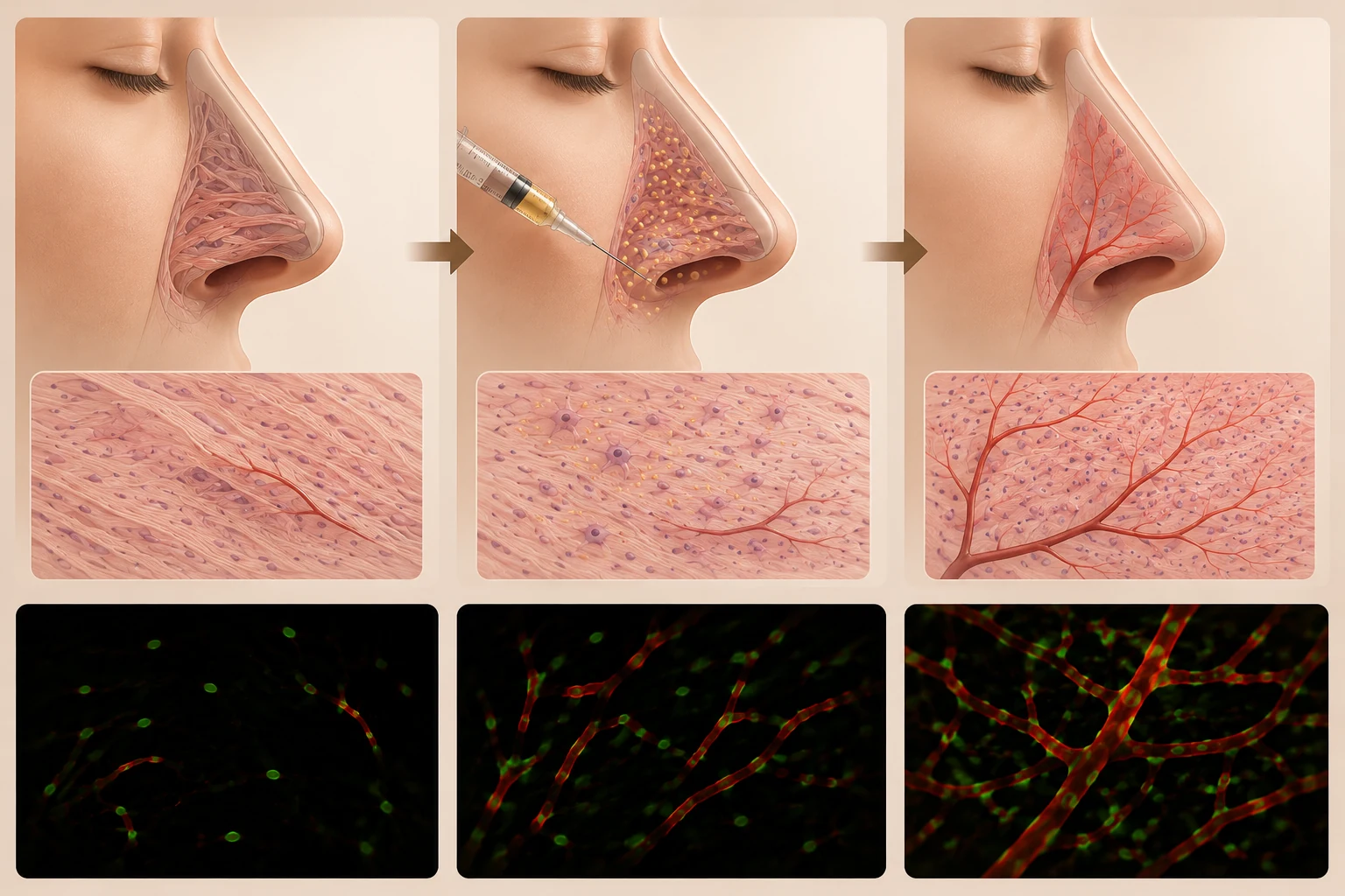
When rhinoplasty is repeated several times, or when a silicone implant causes side effects such as inflammation, skin perforation, or contracture, the skin and soft tissue become stiff and hard to stretch, blood supply becomes deficient, and the risk of soft tissue necrosis increases.
In such cases, no matter how well the surgery lengthens and raises the nasal framework with autologous cartilage, the problem of the soft tissue covering it must be resolved first.
Rhinoplasty using stem cells is the method of first preparing this stiff, damaged soft tissue to become soft and healthy and then performing rhinoplasty. It belongs to the field of "regenerative plastic surgery," which has recently been very actively studied and applied in plastic surgery.
Stem cell rhinoplasty at RESUM Plastic Surgery uses i) SVF (adipose-derived stromal vascular fraction) and ii) PRP/PRF (autologous platelet-rich plasma).
What Is SVF (Adipose-Derived Stromal Vascular Fraction)?
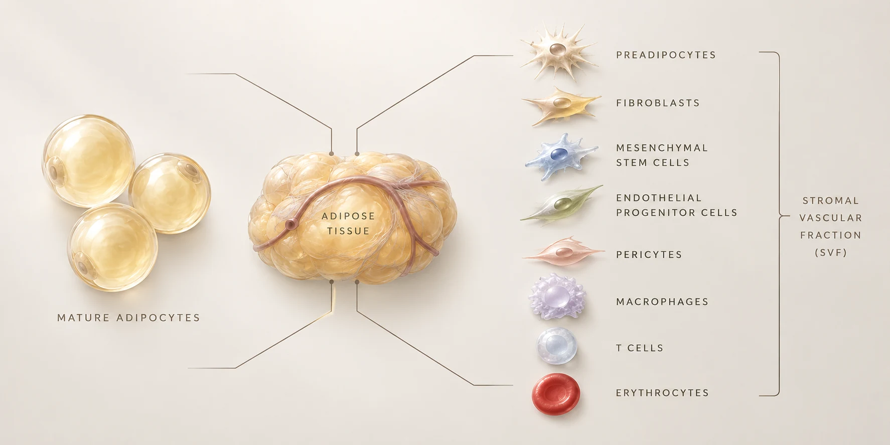
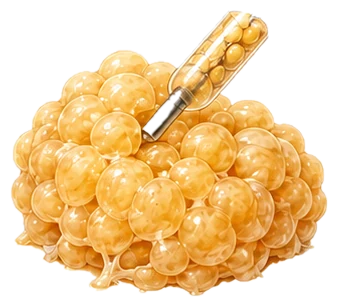
Subcutaneous fat is made up of two main parts.
(1) Mature adipocytes: Large cells that make up most of the volume of fat.
(2) The stromal-vascular compartment between the fat cells: It consists of small blood vessels, pericytes, stem/progenitor cells, fibroblasts, immune cells, and other components.
SVF is therefore what remains, in concentrated form, after fat is processed to remove the large mature adipocytes.
02
Components of SVF
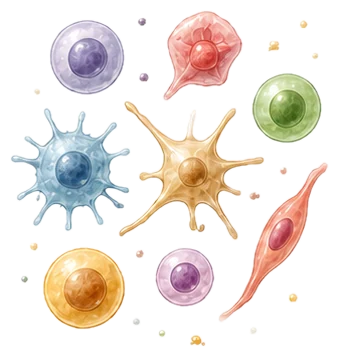
SVF contains not just one type of cell but a combination of several cells essential to tissue regeneration:
ADSCs
(1) Adipose-Derived Stem Cells
(ADSCs): Key cells that can differentiate into cartilage, bone, blood vessels, skin, and other tissues according to their surrounding environment.
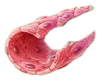
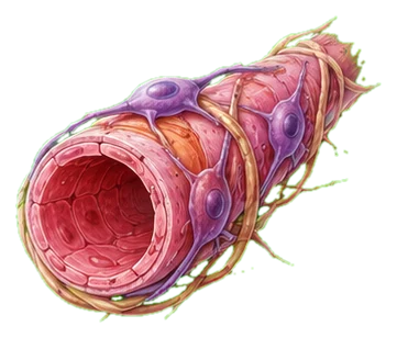
Endothelial & Pericytes
(2) Endothelial Cells and Pericytes
(Endothelial cells & pericytes): They form new microvessels and supply blood circulation.
Immune Cells & Fibroblasts
(3) Immune Cells and Fibroblasts
They heal wounds, regulate inflammation, and build the tissue's supporting scaffold, such as collagen.
Growth Factors
(4) Growth Factors
They play a variety of roles, including tissue regeneration and vascular regeneration.
Why Use SVF?Use in Rhinoplasty
Mechanism of action of SVF in revision surgery and contracted nose correction.
A much stronger tissue regeneration effect appears when various cells and growth factors work together as one team in the form of SVF than when stem cells work alone.
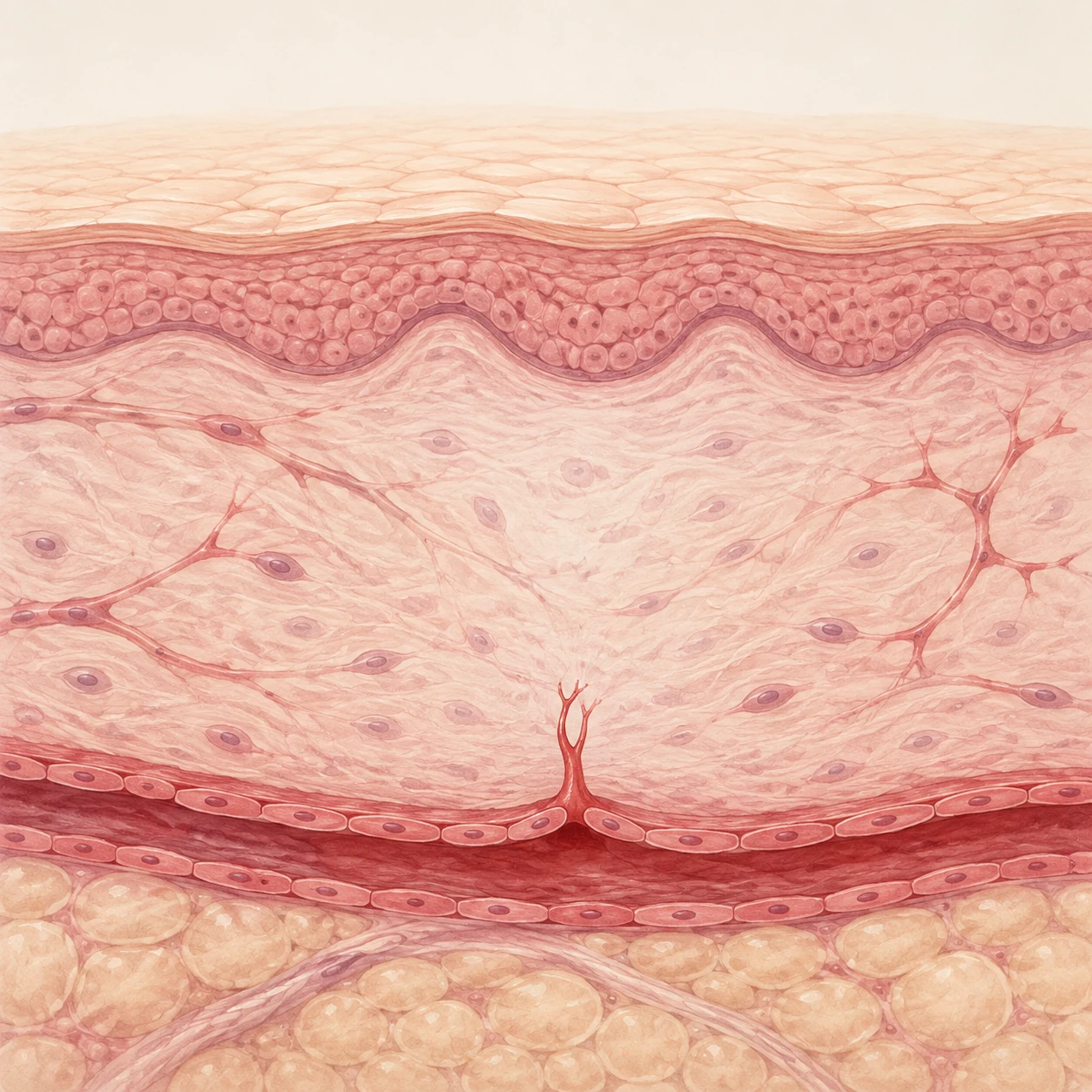
When the nasal skin and soft tissue have hardened and shrunk because of side effects from implants such as silicone or repeated revision surgery, SVF is used to make them soft and elastic and to improve blood circulation, bringing them into a condition suitable for revision surgery.
The greatest challenges in revision surgery and contracted nose surgery are "tightly shrunken skin tissue that resists stretching" and "poor blood circulation in scar tissue."
No matter how attractive and sturdy the internal framework is, if the overlying skin does not stretch or cannot withstand the pressure, it cannot cover the framework and there is a risk of skin necrosis. SVF solves this problem biologically.
Tissue Softening
(1) Tissue Softening and Scar Tissue Relief
The various growth factors and stem cell components in SVF rearrange and break down the structure of excessively accumulated collagen (scar tissue), turning hard nasal skin into a soft, readily stretchable state.
Angiogenesis
(2) New Blood Vessel Formation
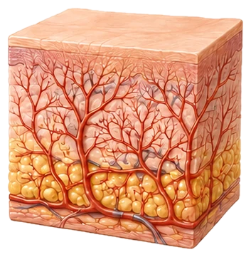
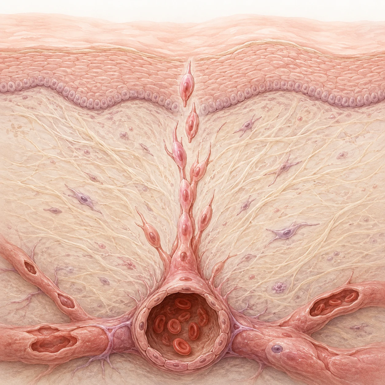
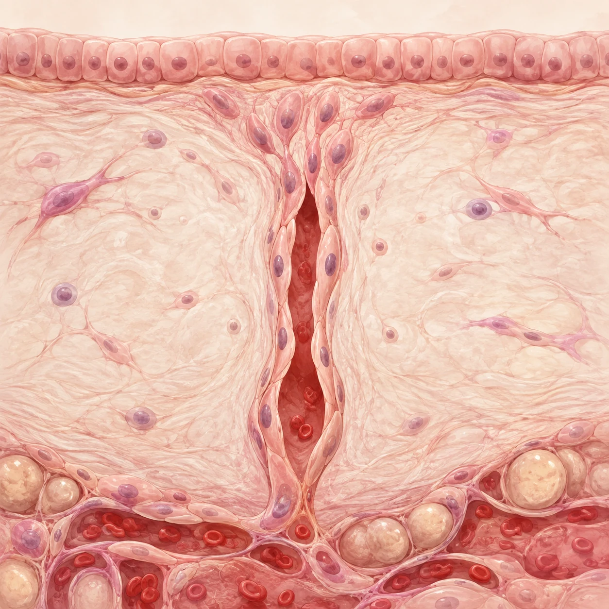
The endothelial cells and pericytes in SVF strongly promote microvessel formation. When blood supply improves, the risk of skin necrosis during revision surgery drops markedly and wounds heal faster.
Anti-inflammation
(3) Anti-Inflammatory Action
Immunomodulatory cells calm chronic foreign body reactions and the inflammatory environment, preventing contracture from progressing further.
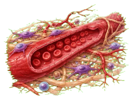
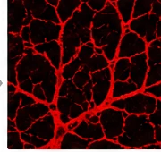
Vascular RegenerationThe Vascular Regeneration Effect of SVF
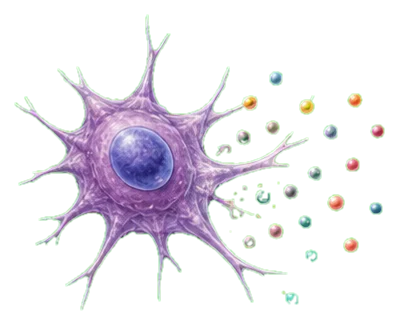
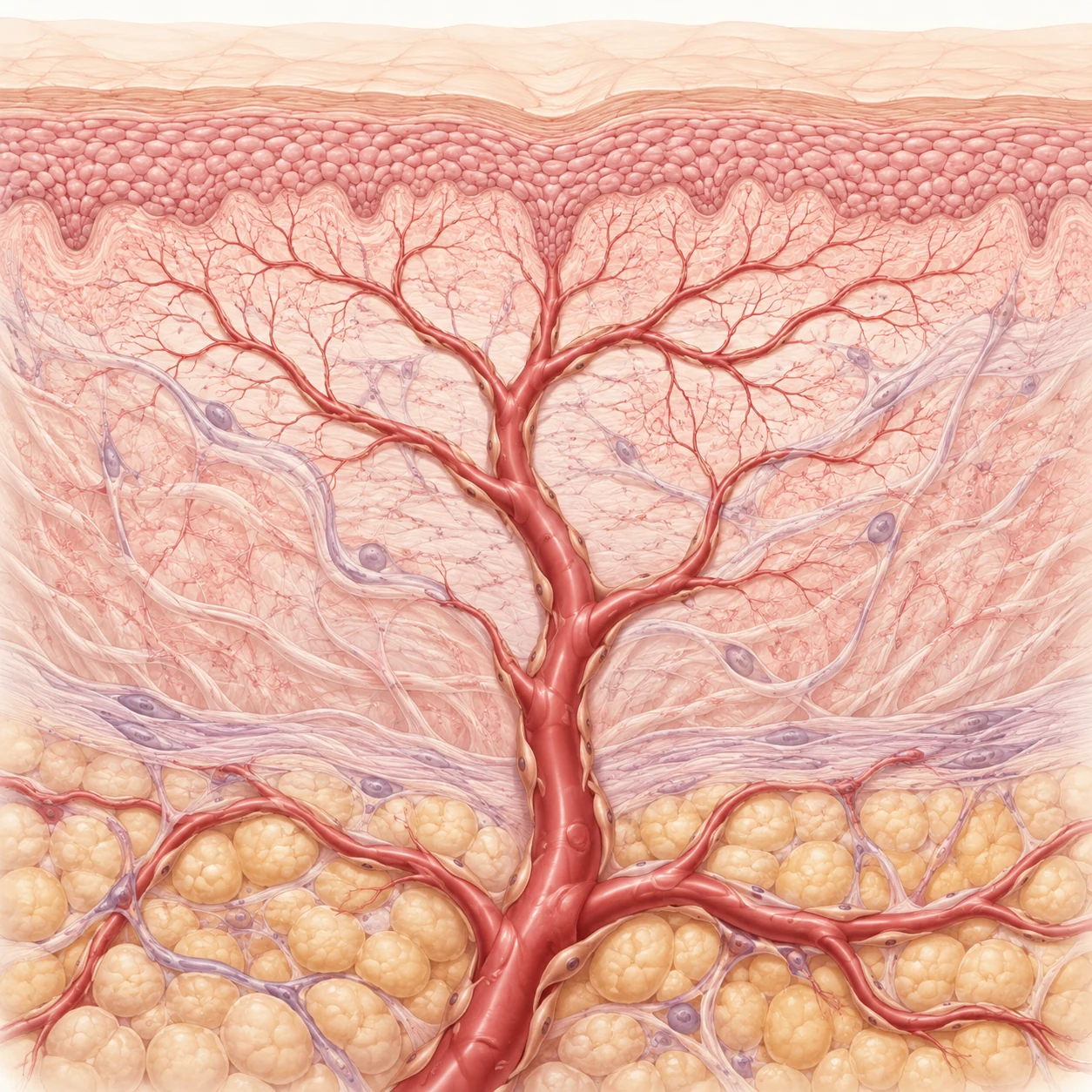
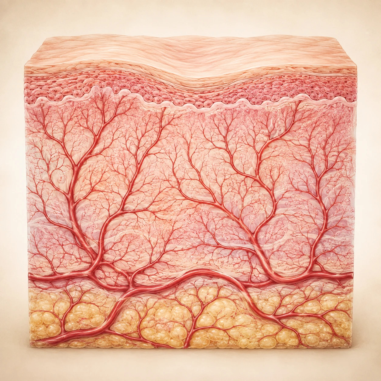
Through the interaction of various cells and paracrine signaling, SVF promotes the formation, maturation, and stabilization of blood vessels.
01
Adipose Tissue Harvesting
(Liposuction)
02
SVF Isolation
(Mechanical/Enzymatic)
03
Cells in SVF
ASC / ADSC (adipose-derived stem cells)
Endothelial cells
Pericytes
Immune cells (macrophages, etc.)
Fibroblasts
04
SVF Injection
05
Vessel Regeneration & Tissue Recovery
Mechanism
Mechanism of SVF Vascular Regeneration
01
Growth Factor Secretion (Paracrine effect)
Secretion of various growth factors activates endothelial cells and promotes angiogenesis
02
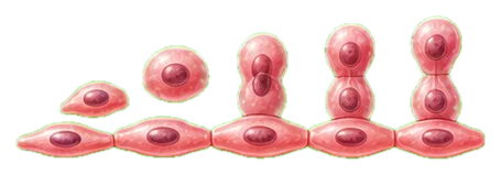
Endothelial Cell Activation and Proliferation
Promotes endothelial cell migration, proliferation, and tube formation
03
Vascular Tube Formation (Tubulogenesis)
Endothelial cells form tube-like structures, building a network of new blood vessels
04
Vessel Maturation and Stabilization
Pericytes surround the vessels to stabilize them, and differentiation into smooth muscle cells strengthens the vessel wall
05
Improved Blood Flow and Tissue Regeneration
Increased blood flow, improved oxygen/nutrient supply, tissue recovery, and improved function
Vascular Regeneration in Ischemic Tissue (Example)
04Promotion of Wound Healing and Tissue Regeneration↑
Through the synergy of various cells and growth factors, SVF induces angiogenesis and vessel stabilization, supporting tissue regeneration and recovery of function.
SVF in Practice 4. How SVF Is Applied
01
Pre-conditioning
(1) Before Surgery - Pre-conditioning
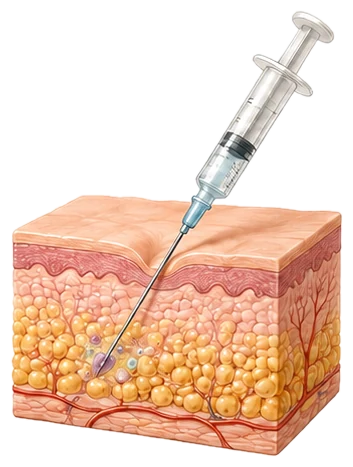
About 1-3 months before surgery, SVF extracted from the patient's own fat is first injected into the skin layer of the nasal tip and bridge, where contracture is severe. This increases the extensibility of the skin, so that when the nasal framework is lengthened during surgery, the skin can stretch safely and provide the space and length needed to cover the framework.
1-3 Months BeforeExtensibilitySkin Space and Length
02
SVF-enhanced Fat Graft
(2) During Surgery - SVF-Enriched Fat Grafting and Dermal Regeneration
After the contracted tissue is dissected and the structure is built with autologous costal cartilage or other materials, autologous fat mixed with SVF (SVF-enhanced fat graft) is grafted, or SVF is combined with artificial dermis and laid over the area, to reinforce the thickness of the thinned, damaged nasal skin.
Compared with conventional fat grafting, mixing in SVF maximizes the survival rate of the fat cells, and it has shown an excellent effect in improving scar quality in areas with poor blood flow, such as the nasal tip (alar zone).
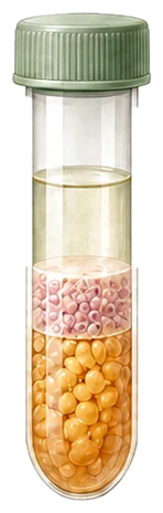
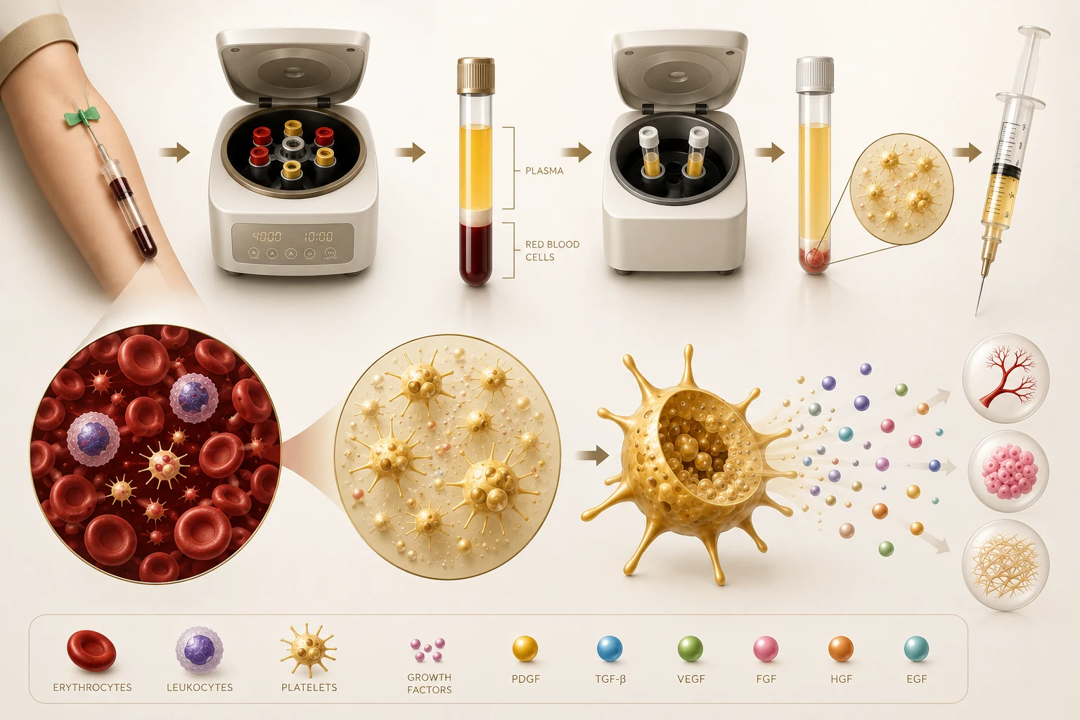
PRP (autologous platelet-rich plasma) is a platelet concentrate made by centrifuging the patient's own blood. It contains numerous growth factors at high concentration and, like SVF, is widely used to "biologically restore the damaged, hardened soft tissue environment of the nose," and its effect is excellent.
After centrifugation, the blood separates roughly into PPP (platelet-poor plasma) at the top, PRP ± buffy coat in the middle layer, and RBCs at the bottom.
02
Components of PRP
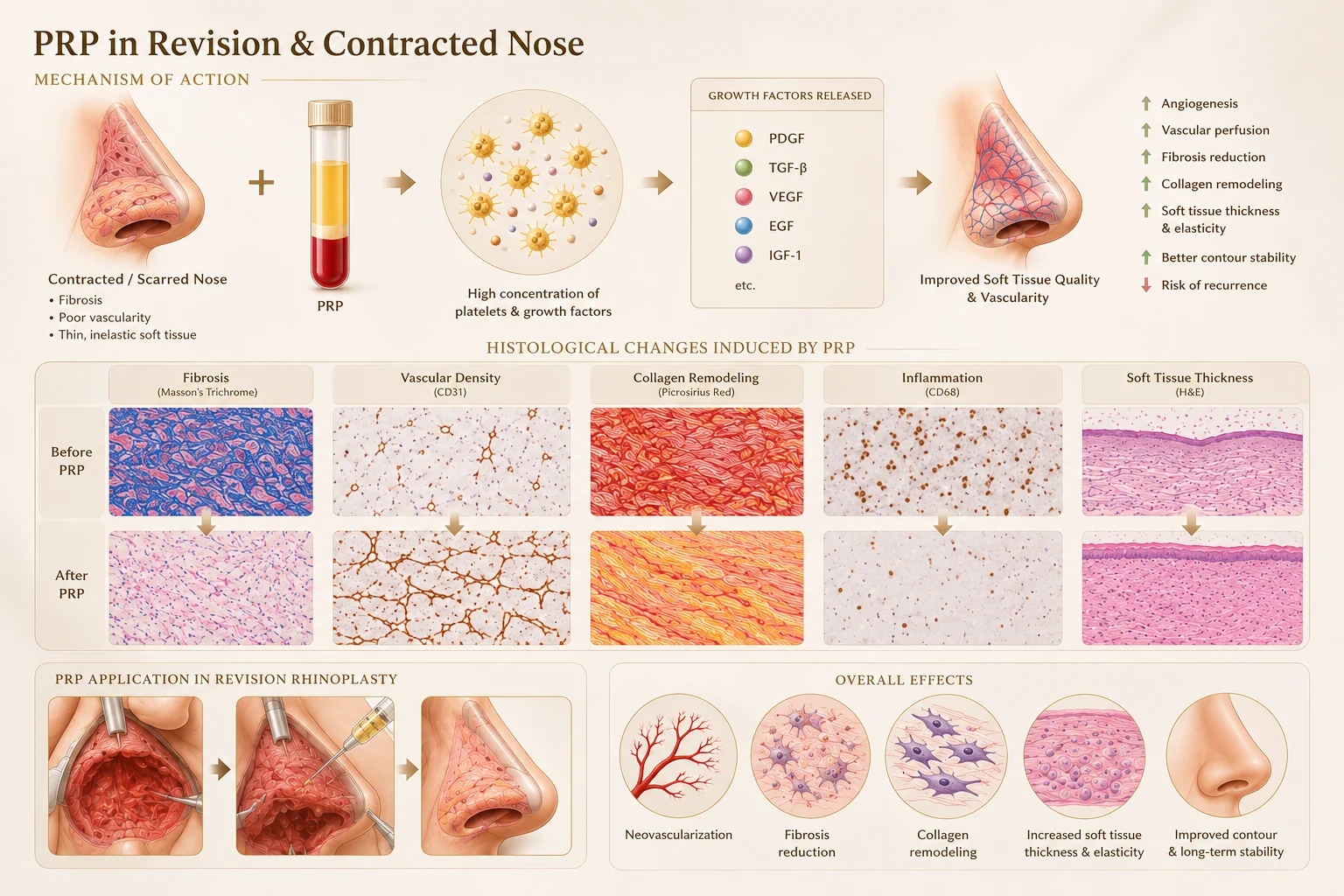
The core of PRP is concentrated platelets, and depending on the preparation method it takes forms such as P-PRP, which is low in leukocytes, and L-PRP, which contains leukocytes. After activation, platelet α-granules and other structures release a variety of bioactive substances, including PDGF, TGF-β, VEGF, EGF, and IGF-1, which are involved in tissue regeneration, angiogenesis, cell proliferation, and ECM remodeling.
Regenerative EffectsUse in Rhinoplasty and Tissue Changes
Angiogenesis
(1) Blood Supply and Induction of New Blood Vessels
Because of repeated surgery and chronic inflammation around a silicone implant, the skin layer of a contracted nose has shrunken blood vessels, is covered in scar tissue, and has very poor blood circulation. Growth factors in PRP such as VEGF and bFGF promote the formation of new microvessels, normalizing the supply of oxygen and nutrients to the surgical site.
Soft Tissue Recovery
(2) Restoring Flexibility and Regenerating the Nasal Skin (Soft Tissue)
Nasal skin that has shrunk from contracture is hard and does not stretch easily. PRP stimulates fibroblasts to rearrange the extracellular matrix (ECM), including collagen and elastin, into a normal structure, softening hardened scar tissue and helping the skin stretch more easily.
Graft Survival
(3) Maximizing Survival of Grafted Tissue (Cartilage, Fat, Dermis)
Revision rhinoplasty often involves grafting substantial amounts of autologous costal cartilage, ear cartilage, acellular dermal matrix, or autologous fat. For these living tissues to survive on a scarred bed and settle quickly, blood vessels must connect to them quickly. Applying PRP around the graft or mixing it with the graft can lower the graft's resorption rate and raise its survival rate.
Combined SynergyDifferences Between SVF and PRP and Their Combined Use
In clinical practice PRP is often used alone, but the "combination cocktail approach" of mixing SVF and PRP together is known to produce the strongest synergy.
PRPSVF
SourceExtracted from the patient's bloodExtracted from the patient's adipose tissue
RoleActs as the "fertilizer (signal)" that wakes and stimulates stem cellsActs as the "seed" that differentiates on its own and builds tissue
CostKRW 500,000 per sessionKRW 4-5 million per session
Synergy Effect
When SVF (the seed) enters the nasal tissue and begins to differentiate into cells, injecting PRP (the fertilizer) along with it rapidly activates the surrounding environment, and the speed of blood vessel formation and tissue regeneration increases dramatically.
Conclusion
In revision rhinoplasty and contracted nose correction, PRP is not used for a merely cosmetic purpose; it is "a biological safeguard that underpins the structural success of revision surgery." If setting the internal support (costal cartilage and the like) firmly in place is the carpenter's job, PRP enriches the soil (the skin tissue) surrounding that support, helping the surgical result hold over the long term without warping.
Case Study9 Surgeries in All: A Journey of Reconstruction Begun Anew
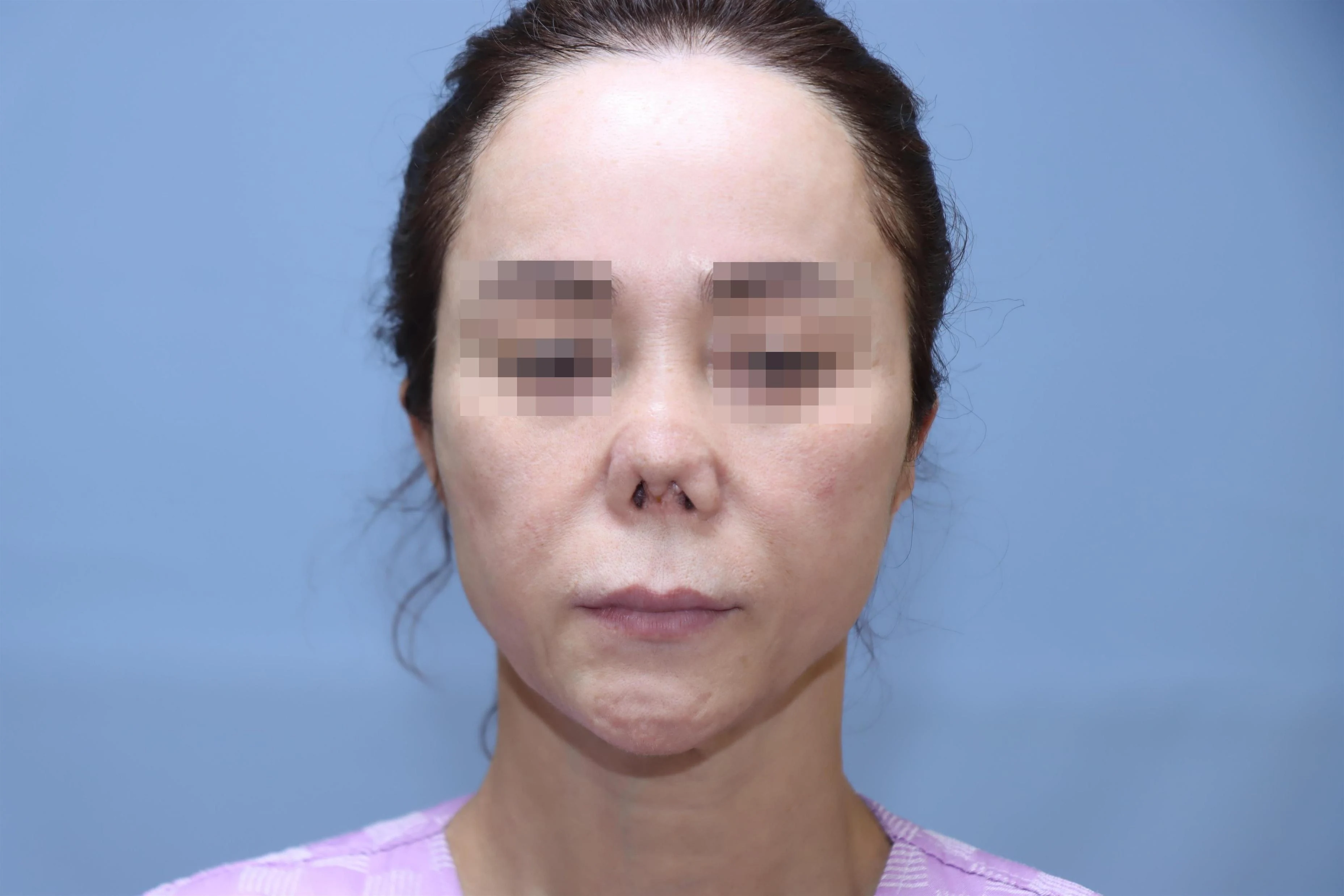
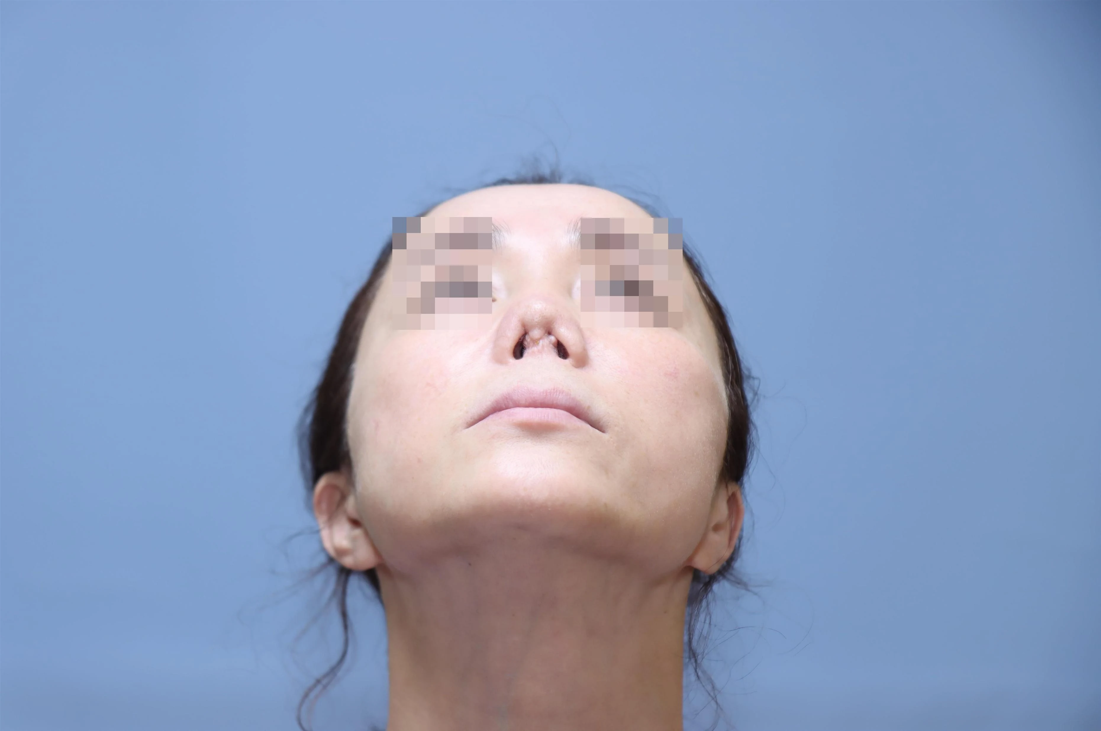
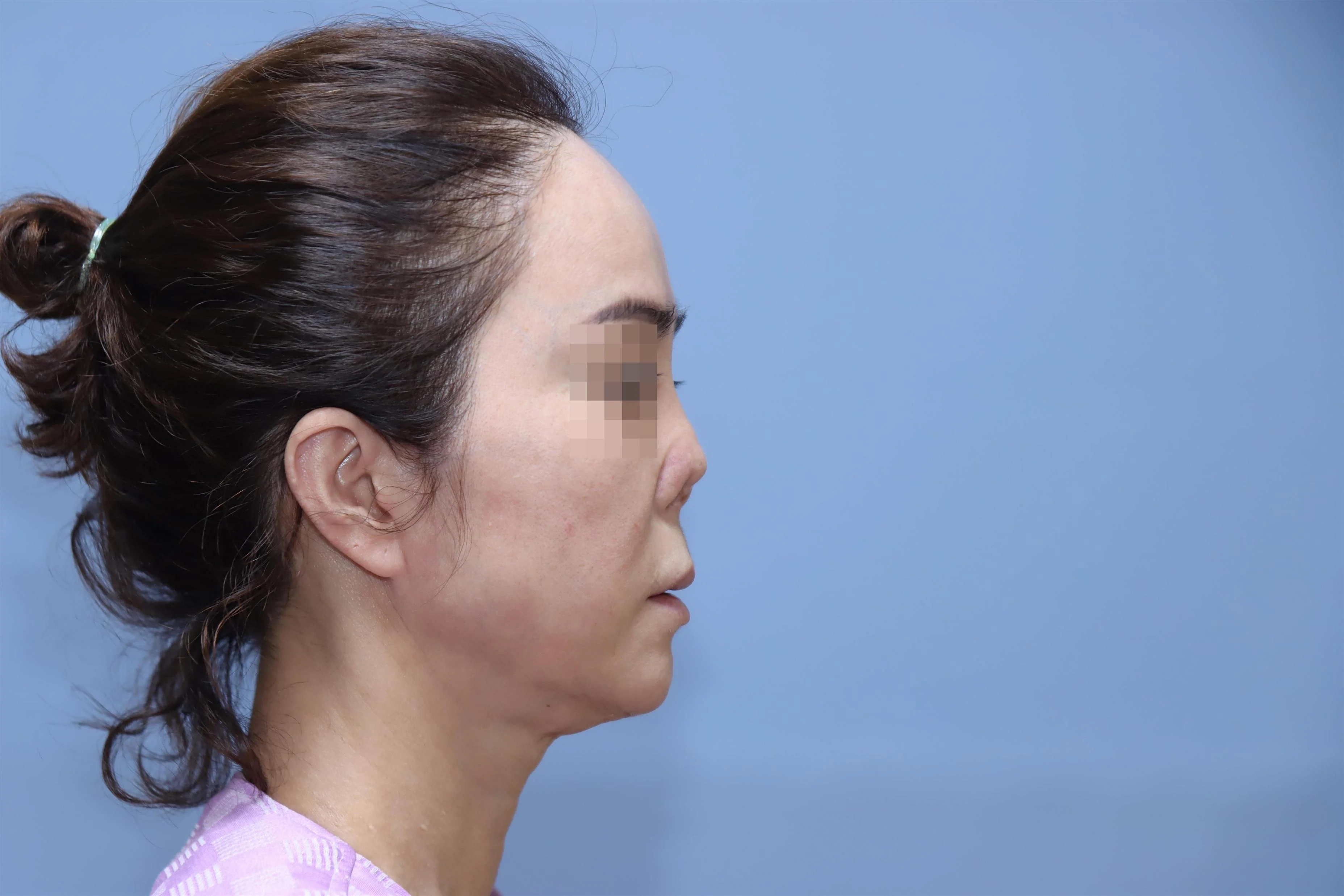
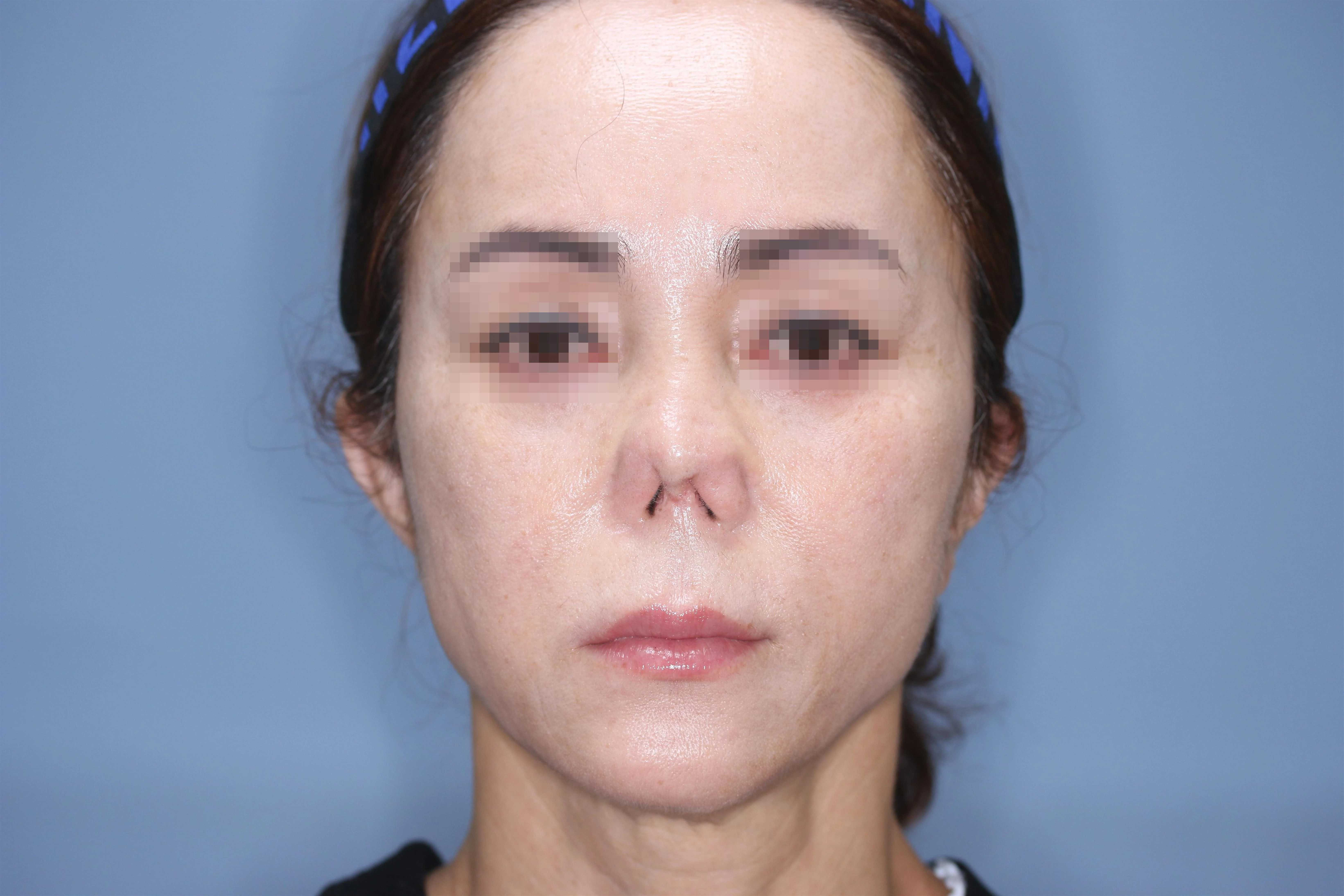
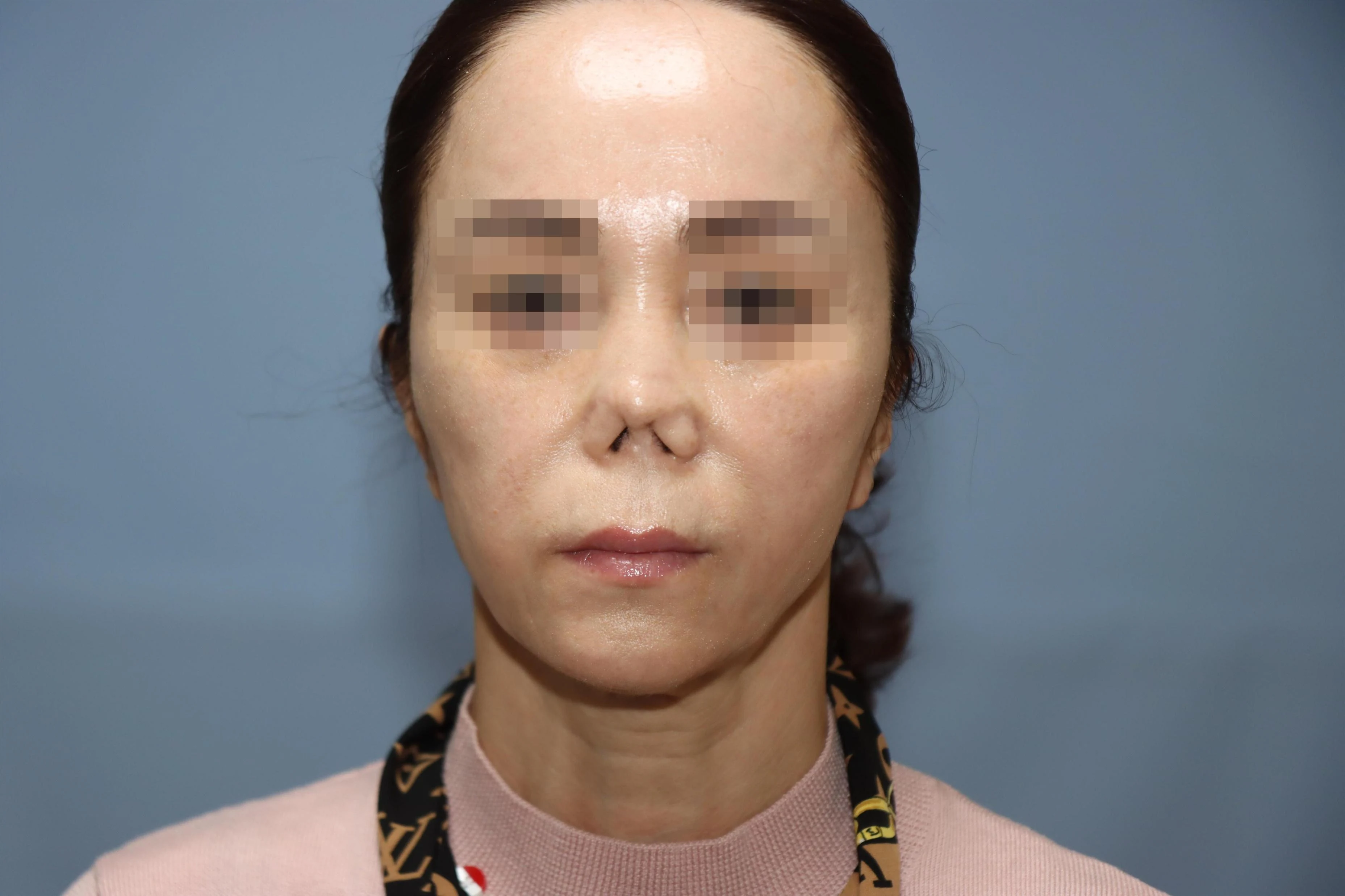
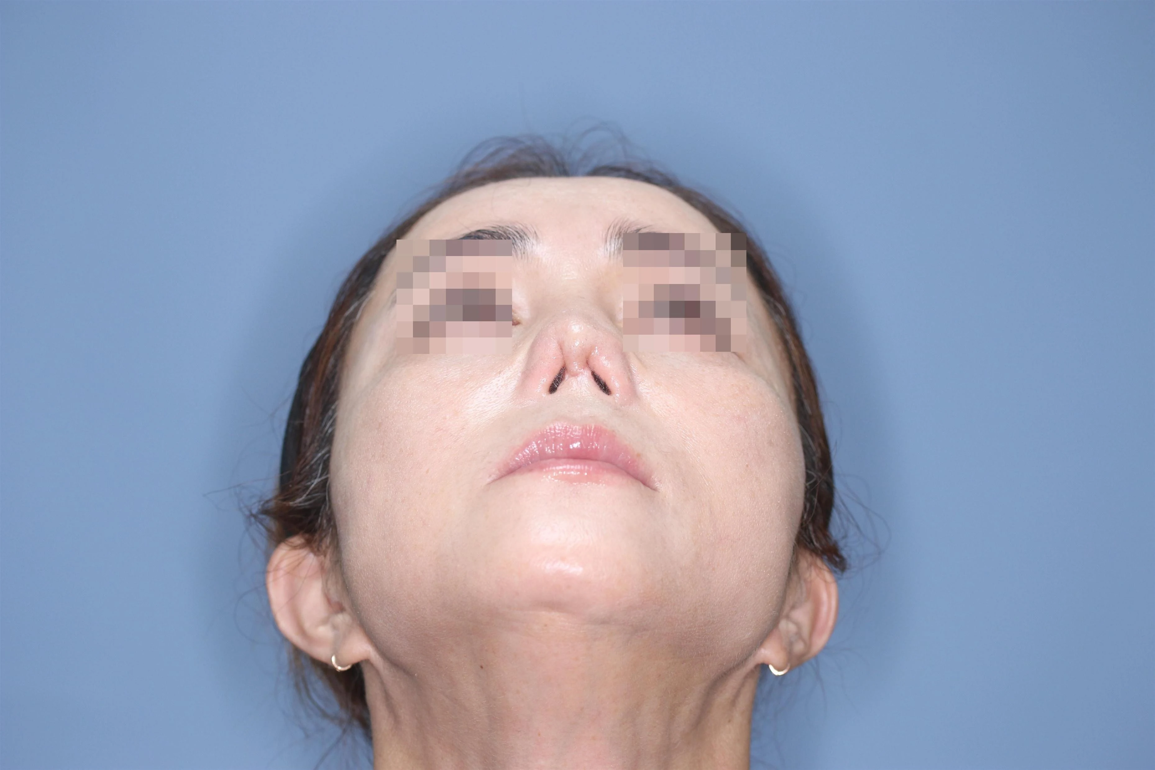
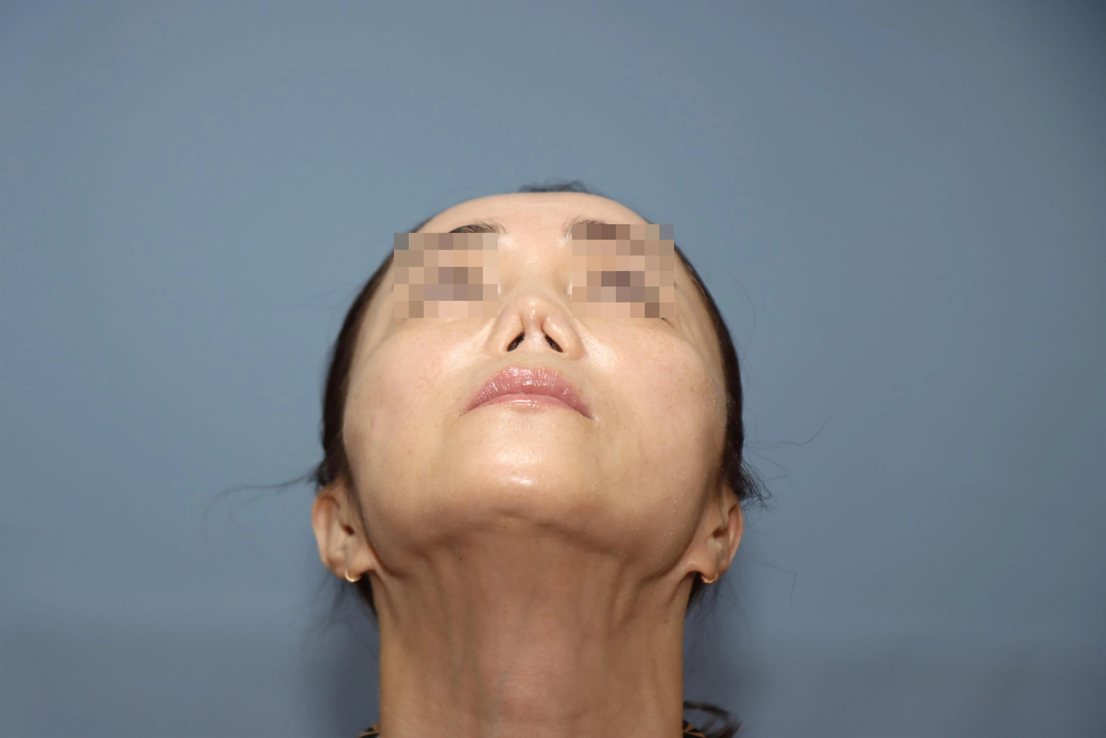
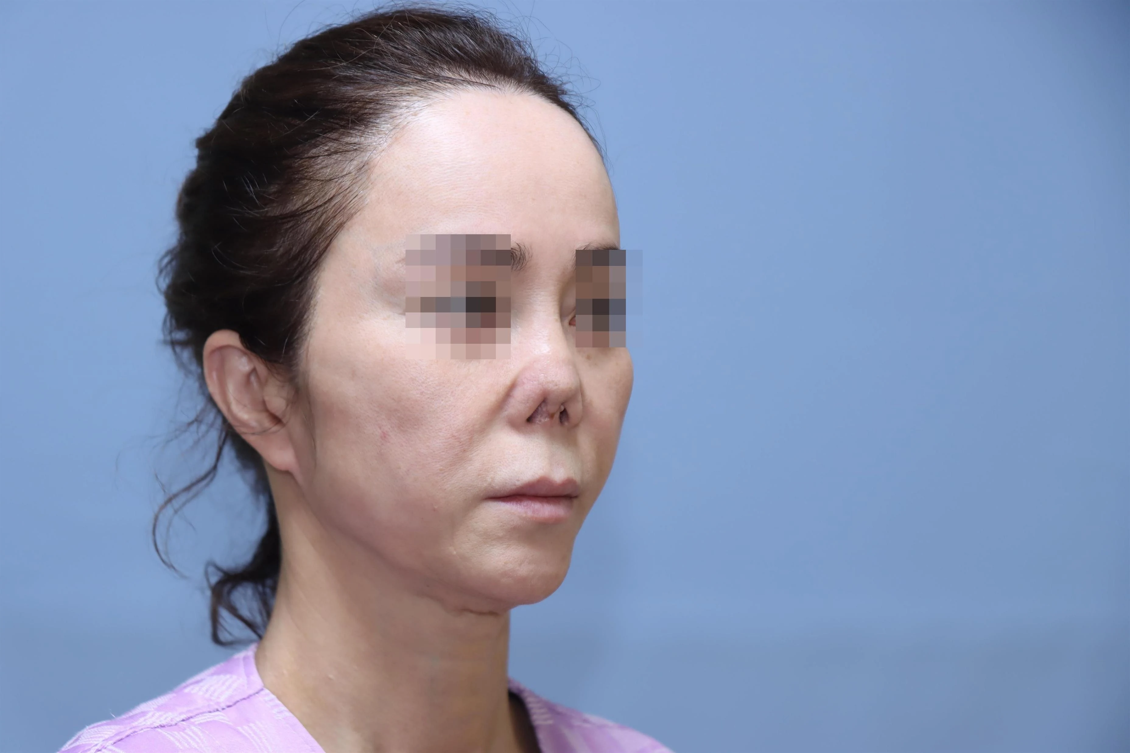
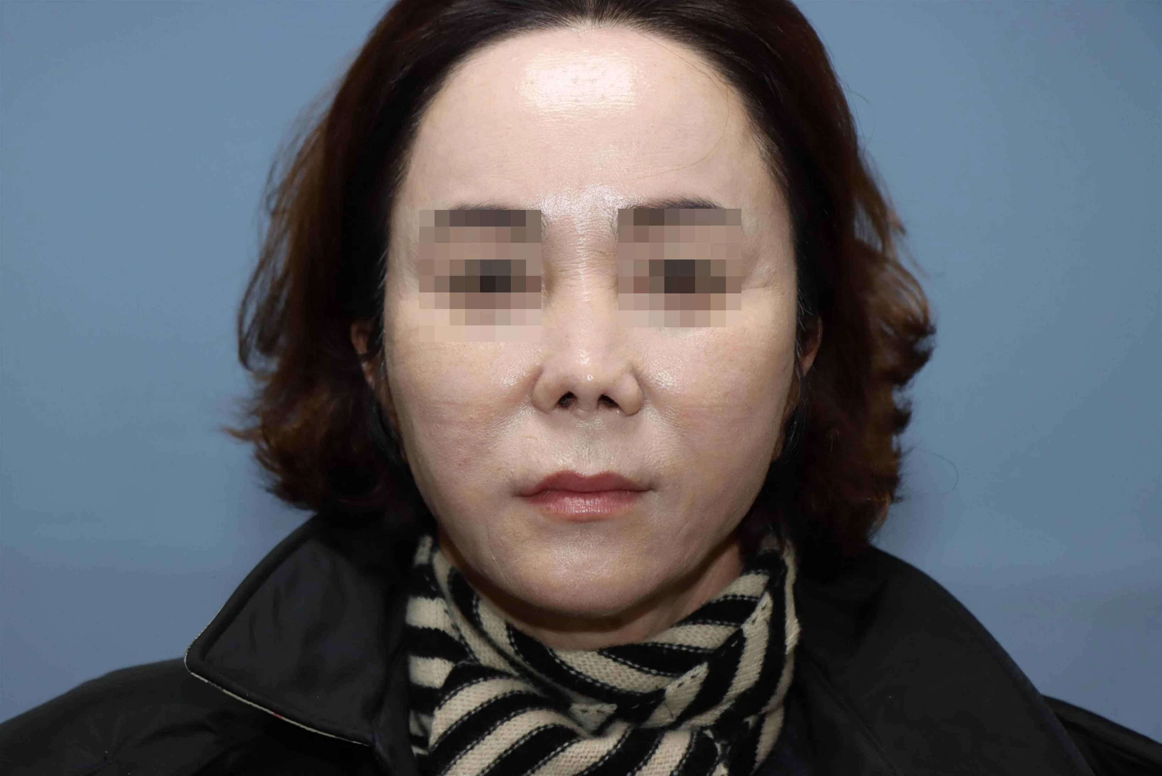
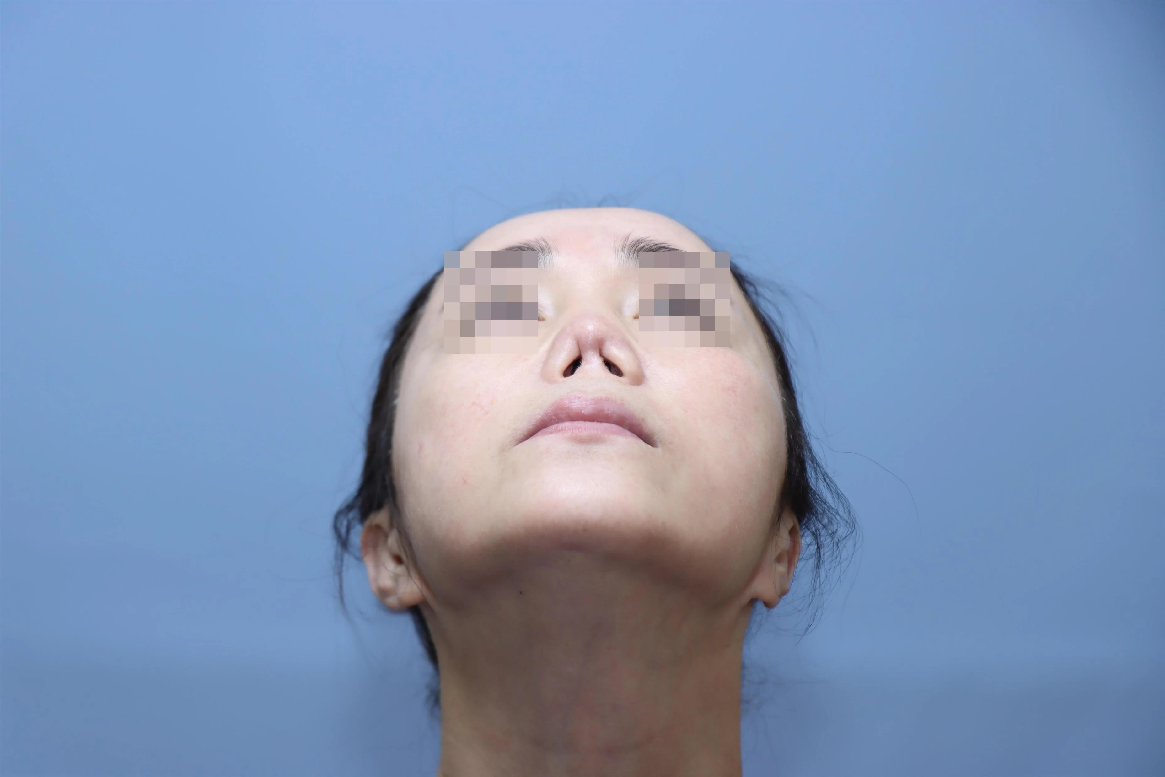
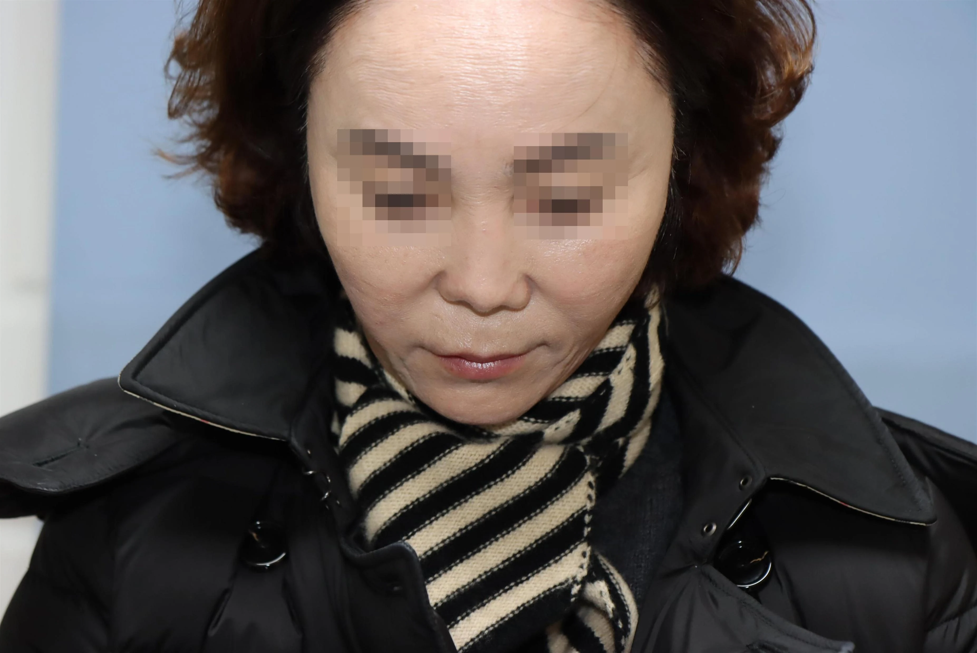
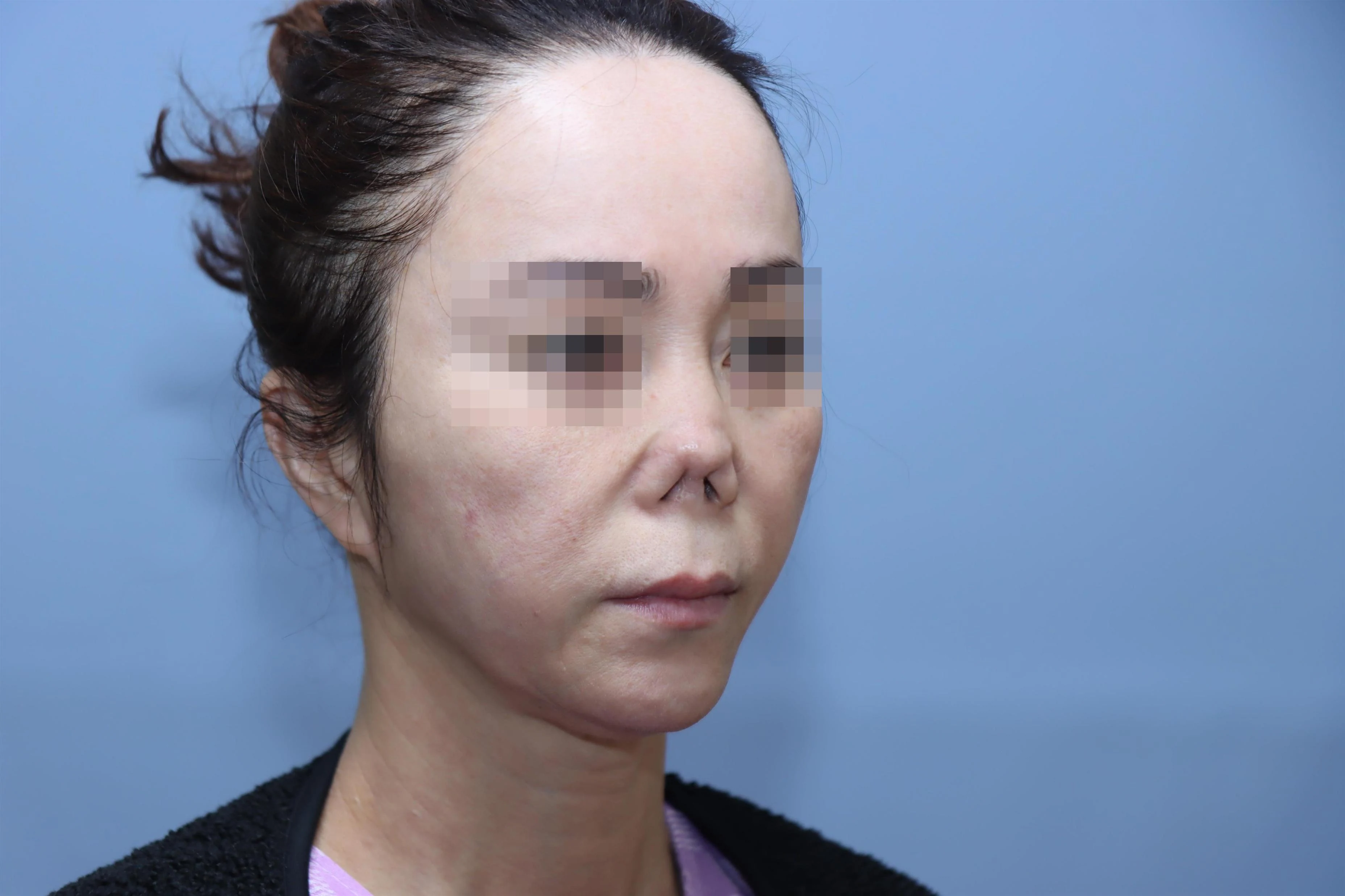
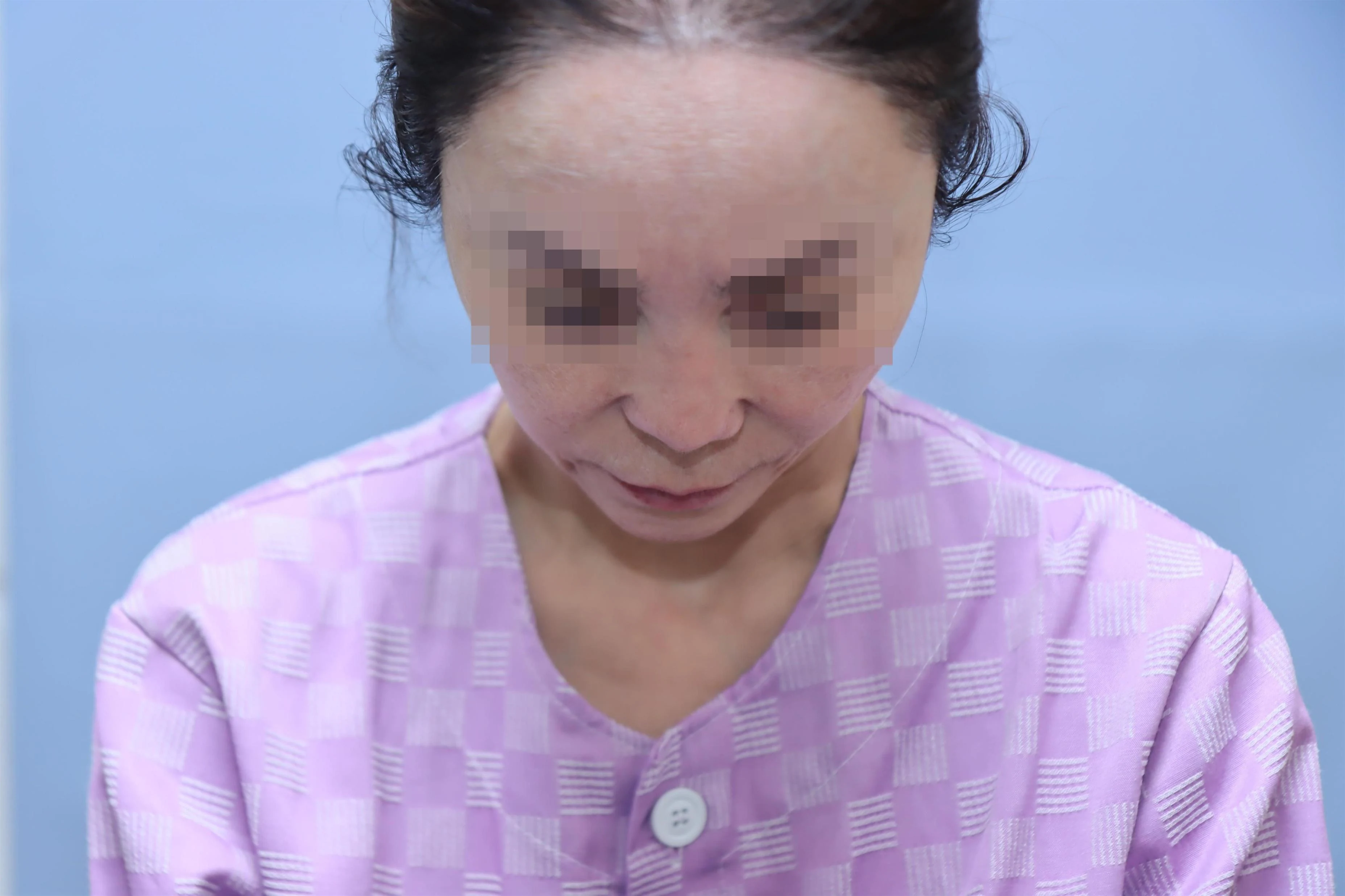
When this patient first came to our clinic several years ago, the patient had already undergone 3 surgeries at other clinics. There was already soft tissue deformity and contracture at that time, so we operated only after making sure the patient understood that this was to be the last surgery. After the surgery the patient was satisfied and thanked us, and we heard nothing further until, one day, the patient reappeared looking like this.
When we asked what had happened, the patient hesitated, kept apologizing, and then tearfully explained. After the surgery at our clinic, the patient had at first been very happy and was often told the nose looked pretty, but at some point began, little by little, to want more: a nose just a bit higher and longer. We had advised that it was better to stop there, and the patient hesitated, but after much thought went to another clinic and underwent augmentation rhinoplasty twice. Infection developed both times, and the patient came to us with the nose badly distorted and discharging pus.
PH
After revision surgery at our clinic in February 2017, the patient was satisfied at first but wanted more nasal length and a higher bridge, and so went to another clinic and underwent augmentation rhinoplasty twice with autologous costal cartilage and acellular dermal matrix. Infection developed both times, and the acellular dermal matrix was removed. January 2021: surgery with acellular dermal matrix & autologous costal cartilage → infection developed. Removed 20 days later. Contracture developed after removal. May 27, 2022: augmentation rhinoplasty with acellular dermal matrix alone → infection again. Acellular dermal matrix removed 5 days later. The inside of the nose was irrigated 2 times, but infection was still present.
P/E
Infection was still present, contracture was severe, and the nasal tip was also badly deformed.
Plan
We decided to perform infection removal surgery first and then, after 1 year, reconstructive surgery with autologous costal cartilage.
Infection RemovalSecond Surgery at Our Clinic, 7th Overall
Using an open approach, we removed all of the infected tissue. Facing a 7th surgery brought on by the patient's own desire for more, the patient was very depressed and struggling, but was too afraid to go to any other clinic and so, apologetically, had come back to ours, promising to follow our instructions faithfully from now on. We decided to divide the removal and the reconstruction into two separate surgeries, and performed the removal surgery first.
During surgery we found that a silicone capsule, which had been removed once before, along with costal cartilage, septal cartilage, ear cartilage, and granulomas, remained inside the nose, and we removed them all thoroughly. Afterward, skin contraction caused the columella to retract further and the nostrils to narrow, and the skin was stiff and uneven. To prepare for reconstructive surgery, the patient then received PRP 3 times at 2-week intervals, starting 1 month after surgery. At 5 months after surgery, following 3 sessions of PRP injection therapy, the contraction had eased and the tissue was somewhat softer than at 1 month, but the condition was still not sufficient for reconstructive surgery.
Before Removal
1 Month After Infection Removal
5 Months After Infection Removal
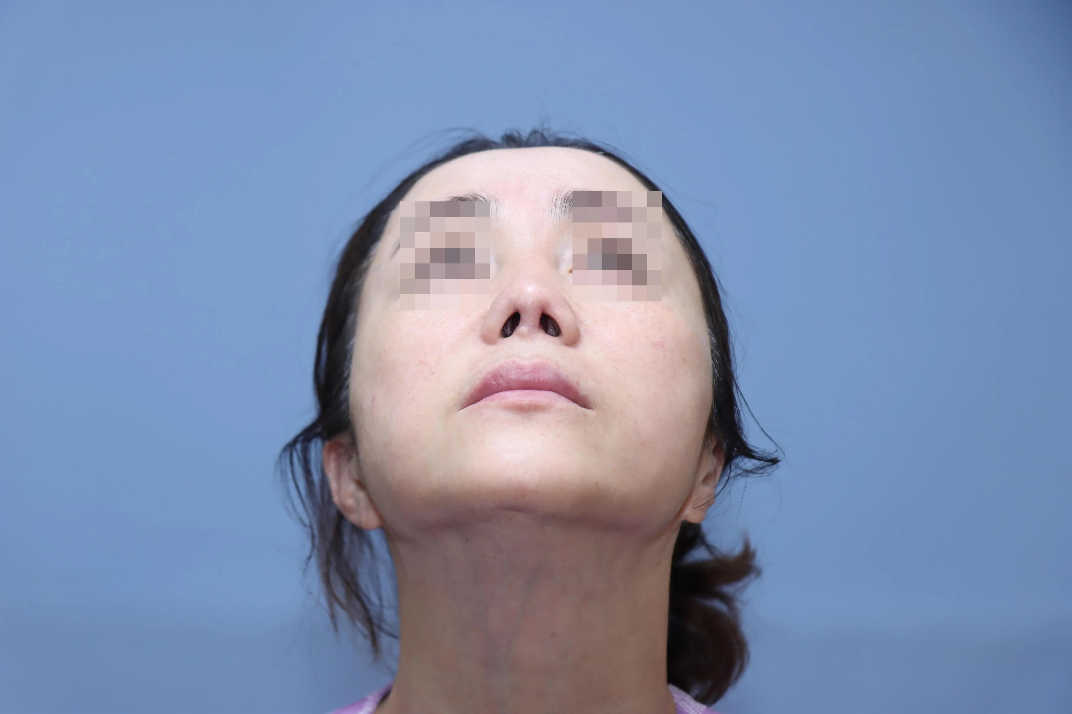
Before the removal surgery, granulomas clung to the columella as small bead-like lumps; after surgery they were cleanly removed and the swelling subsided markedly. At 1 month after the removal surgery, the skin and cartilage of the nasal tip had shrunk from contracture and appeared pinched and notched, and swelling and scar tissue on the inner side of the columella were visible. In the photo at 5 months after removal, the swelling has gone down and the soft tissue can be seen to be somewhat softer and smoother.
Before Removal Surgery
1 Month After Removal
5 Months After Removal
Before the removal surgery the nasal tip was discolored red; after removal the skin color also returned. Between 1 month and 5 months after removal, there is a difference in the maturation of the skin and soft tissue.
Before Removal
1 Month After Removal
5 Months After Removal
At 1 month after removal, contraction was severe, the nose was more upturned, and the retraction of the columella had also worsened; at 5 months, after the autologous blood-derived stem cell treatment, the skin and soft tissue had become markedly softer and smoother.
Before Removal
1 Month After Removal
5 Months After Removal
First Nasal ReconstructionFebruary 2023: Third Surgery at Our Clinic, 8th Overall
After the infection removal surgery, the patient waited 1 year, longing for the day reconstructive surgery would be possible. Because the nasal skin and soft tissue were so severely contracted and stiff, 1 year was the minimum waiting period needed. However, functionally nasal breathing was difficult and outwardly the deformity was severe, so the patient's social life and daily life were greatly affected, and the patient was desperate to have the reconstruction even one day sooner. We therefore performed autologous blood-derived stem cell (PRP) treatment 3 times at 3-4 week intervals and agreed to carry out reconstructive surgery 8 months after the removal surgery.
However, because the damage and contraction of the framework and soft tissue were so severe and the skin was not sufficiently soft and healthy, the nose might not lengthen as much as desired; the patient understood this and agreed.
CC
The nasal tip was extremely shrunken and the nose had become shorter. The bridge was low.
P/E
The septum had collapsed and was deviated, and the nasal cavity was so narrow that even an endoscope could not be inserted. The nasal tip and alae had shrunk, with severe pinching and notching, and the columella was also retracted inward.
OP
We built an L-strut septal framework with autologous costal cartilage to lengthen the nose, and corrected the tip pinching and notching by opening out the alae with alar cartilage grafts (LCSG). On the dorsum, we performed augmentation with acellular dermal matrix.
Before Reconstructive Surgery
Before Reconstructive Surgery
As shown, after reconstructive surgery the pinching and notching of the nasal tip improved and the alae opened out; as a result the external nasal valve widened and nasal breathing returned to normal. The nose was not as long as hoped but had lengthened considerably, and the SSTE (skin and soft tissue) over the dorsum was in better condition than expected, smooth and free of irregularities.
Before Surgery
10 Months After Reconstructive Surgery
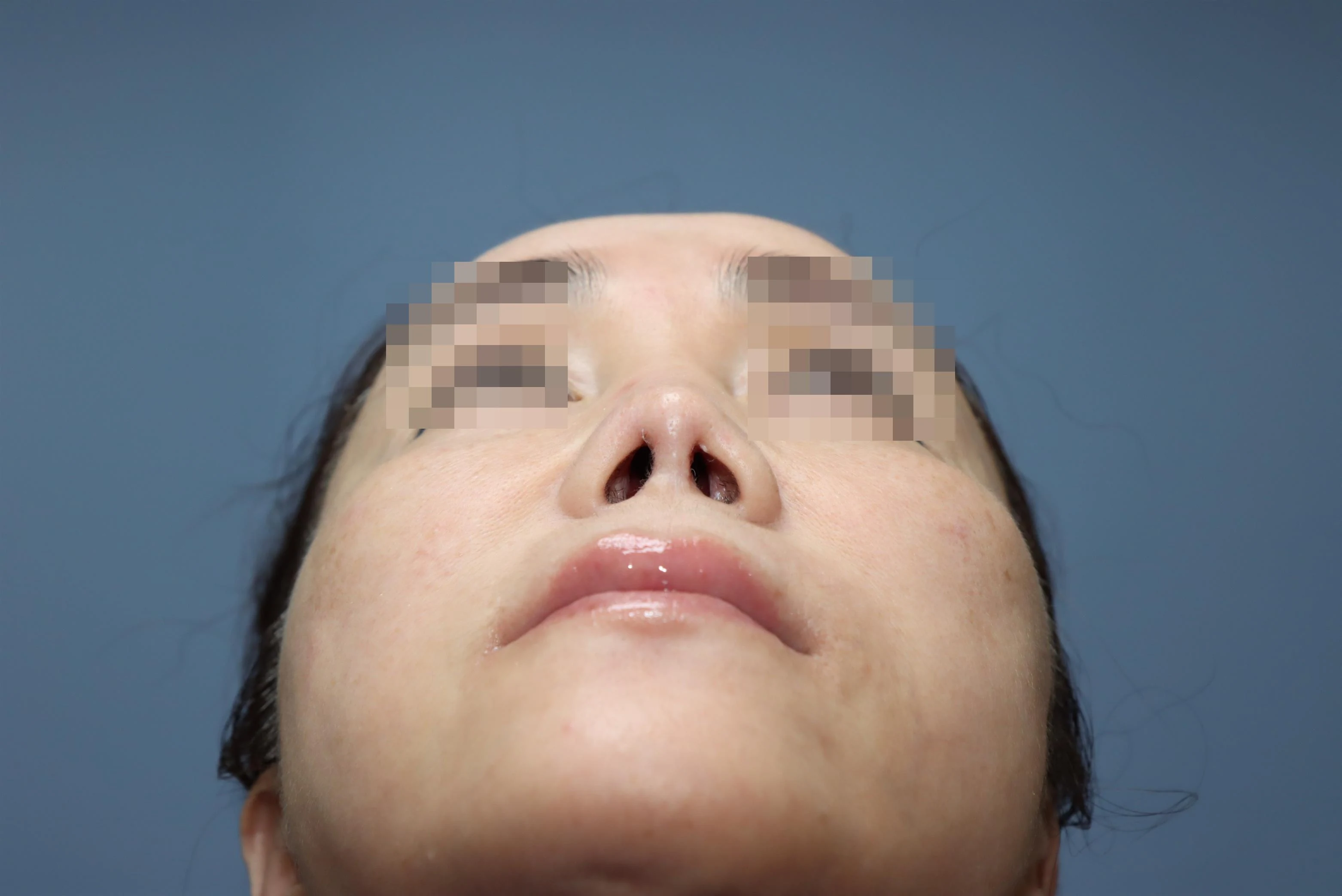
The pinching and notching of the nasal tip improved markedly after surgery, and the size of the nostrils also returned almost to normal.
Before Surgery
After Reconstruction
After reconstructive surgery the alae opened out well, and the nasal tip and columella projected naturally.
Before Surgery
After Reconstruction
After reconstructive surgery, the dorsal line and height, the shape of the nasal tip, and the position of the columella came close to those of a normal nose. The length of the nose left a little to be desired, but because the skin had been so stiff and in such short supply, the patient was also satisfied, considering it as good a result as could be achieved.
Before Surgery
10 Months After Reconstructive Surgery
The columella, which had been bent and retracted, was straightened and projected well after surgery, and the skin also looks healthy apart from being somewhat thinner.
Before Surgery
After Reconstruction
Skin and Soft Tissue TreatmentSoft Tissue Changes After PRP Treatment
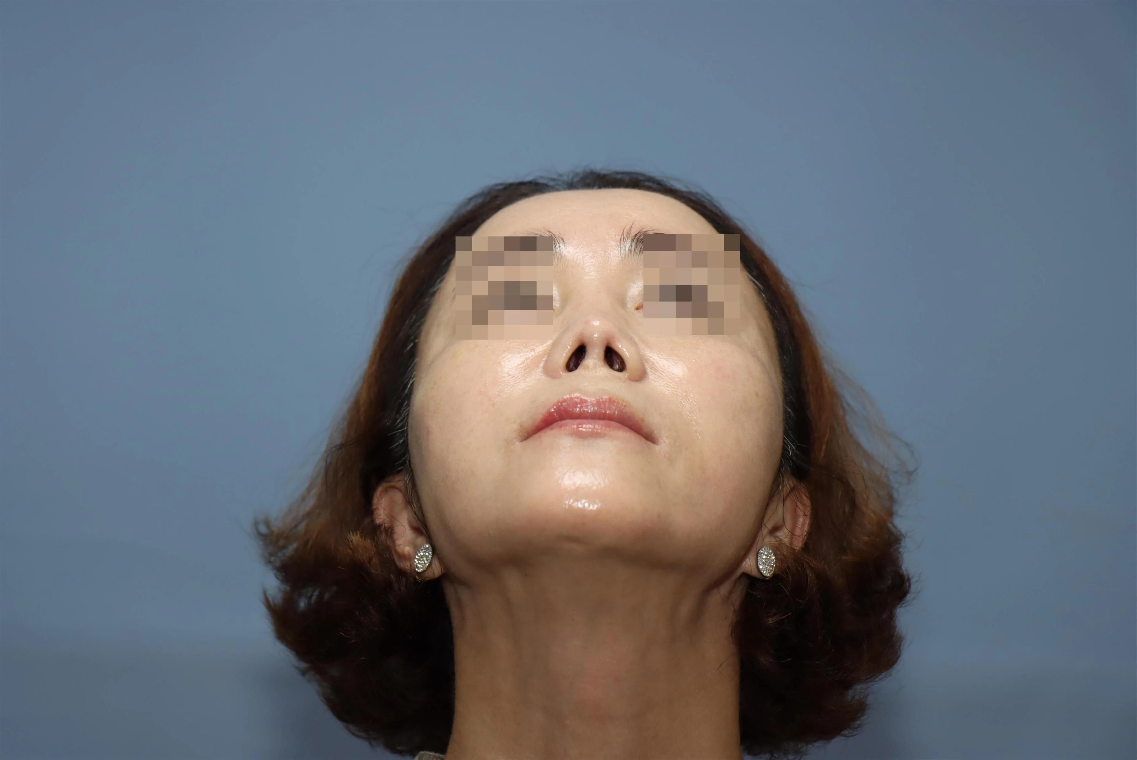
Starting 3 months after surgery, autologous blood-derived stem cell (PRP) treatment was performed 3 times at 3-4 week intervals to make the SSTE (skin and soft tissue) healthy and soft. As a result, the skin and soft tissue gradually became thicker, more supple, and more elastic. By 2 years after reconstructive surgery, the skin and soft tissue had become very soft.
3 Months After the 1st Reconstruction
8 Months After the 1st Reconstruction
2 Years After the 1st Reconstruction
As the skin and soft tissue slowly improved, the patient began, little by little, to want more: just a bit more length and height for the nose. At the same time, a new 2nd reconstructive surgery would be the 9th surgery overall, and the patient felt considerable anxiety and fear about whether the soft tissue could withstand it.
3 Months
8 Months
2 Years
What was clear was that after PRP treatment the soft tissue had become much softer and had gained elasticity.
3 Months After the 1st Reconstruction
8 Months After the 1st Reconstruction
2 Years After the 1st Reconstruction
After 2 years the condition of the skin and soft tissue had almost normalized, and the patient, resolved to place one last hope in Dr. Kook, came to discuss a 2nd reconstructive surgery. Things were already much better, the patient said, but as a final step asked us to improve the length of the nose and the deformity of the nasal tip.
In truth, there had been so many surgeries, and the columella in particular carried so much scarring that there was a risk of necrosis; it was a heavy burden, and my feeling was "let this cup pass from me." But the patient was so earnest, pleading to erase cleanly the painful history of rhinoplasty that had resulted from failure born of excessive desire, and to live from now on with a nose that no one would know had been operated on. I could not turn away from a relationship built up over 8 years, and so, truly feeling as though I were taking up a poisoned chalice, I decided to prepare thoroughly and perform the 2nd reconstructive surgery (the 9th overall).
3 Months
8 Months
2 Years
Final Nasal ReconstructionAfter 3 Sessions of Autologous Stem Cell Treatment (SVF): the 9th Surgery Overall (the Final Surgery)
Because the condition of the soft tissue is critical for a final surgery, we decided to perform autologous stem cell treatment (SVF injection) 1 time before surgery and then carry out the final surgery, the 9th, using costal cartilage and perichondrium.
CC
Upturned nasal tip; tip pinching and notching because the tip had not fully opened out; alar rim retraction; and nostril asymmetry (alar rim retraction, notching, asymmetric nostril, nostril show (Lt>Rt)).
OP
The 2-layer autologous costal cartilage spreader grafts and the CS were removed. Autologous costal cartilage was newly harvested and a new L-strut was made to extend the length. Because no alar cartilage remained, alar grafting was performed with costal perichondrium + autologous costal cartilage. Augmentation was performed with costal perichondrium, a cartilage graft (batten graft) of autologous costal cartilage was placed toward the left external nasal valve, and a composite graft was placed on the inner side of the right ala.
Appearance Before Surgery
Appearance Before Surgery
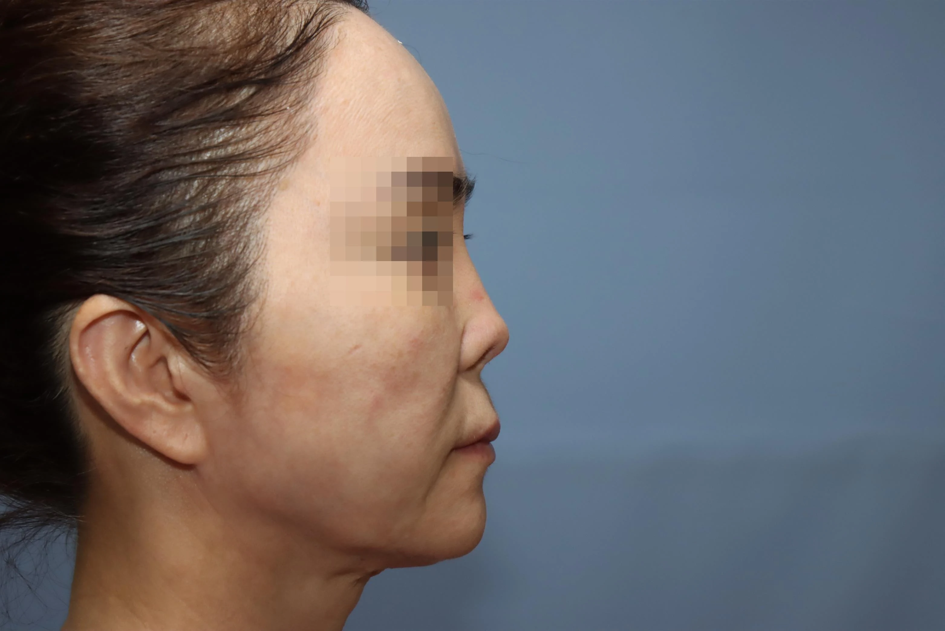
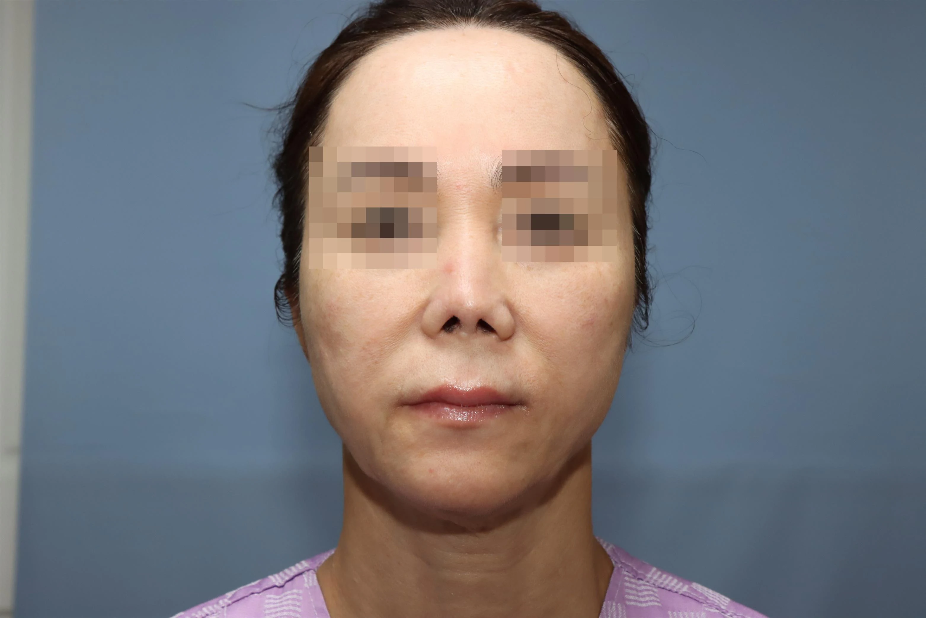
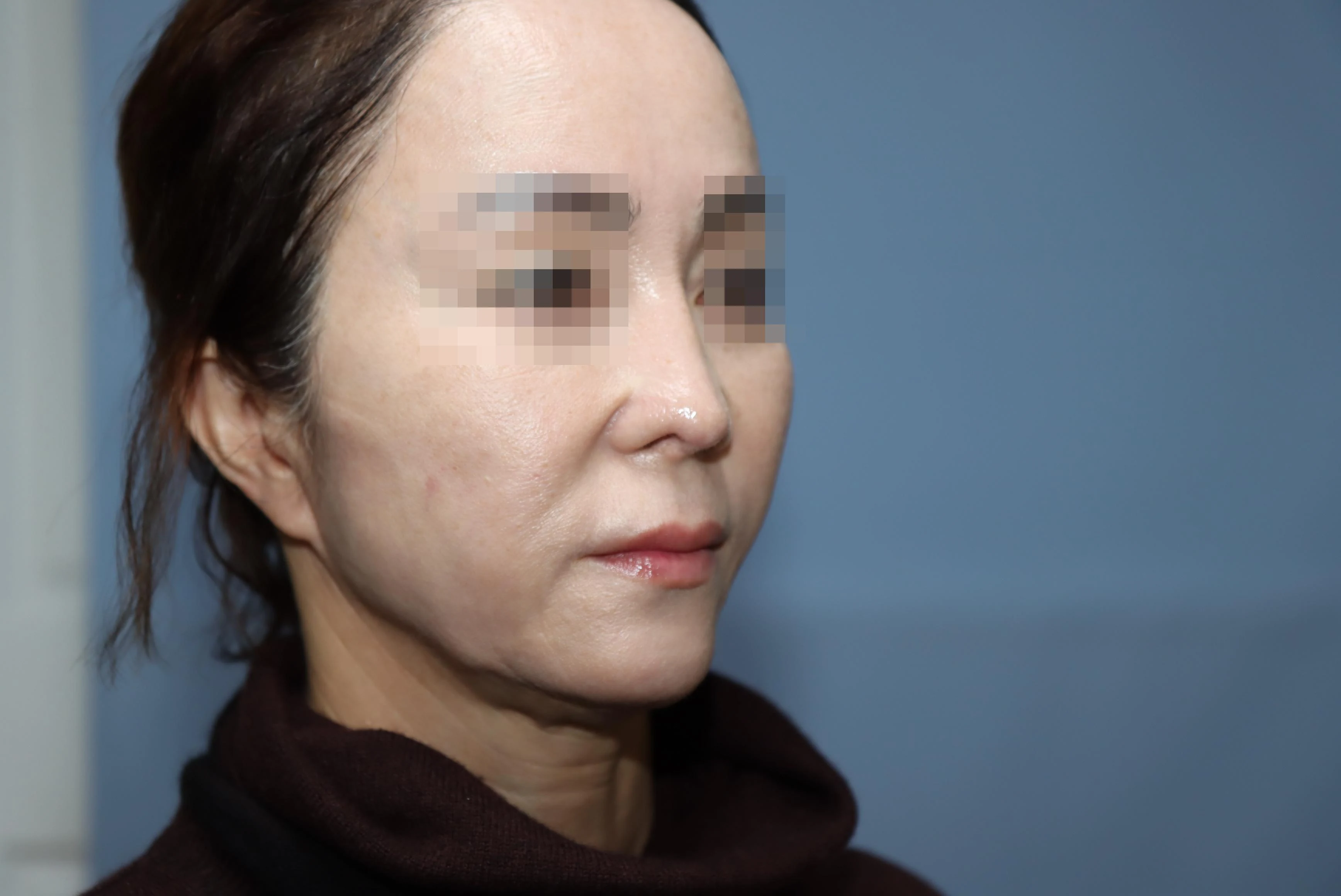
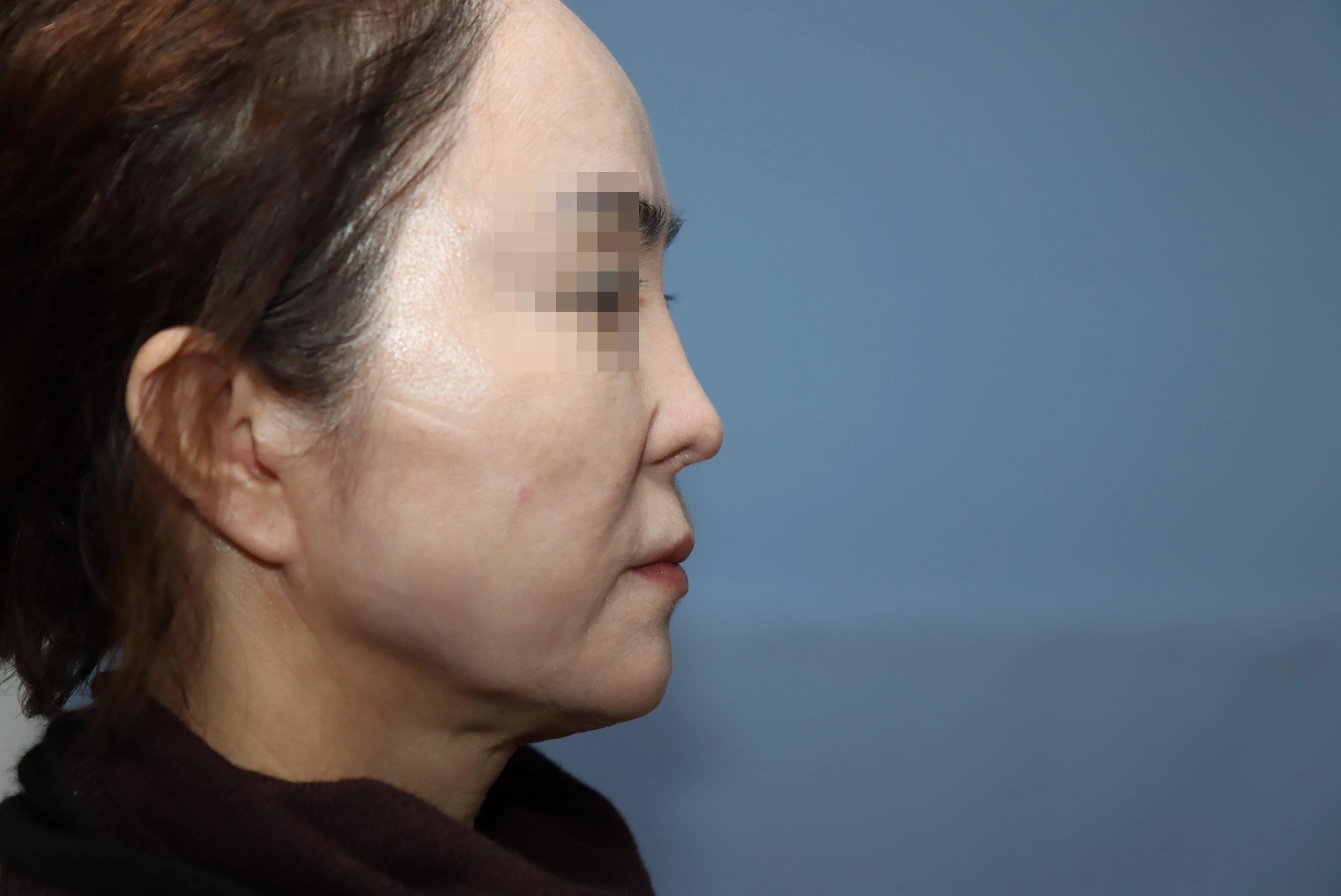
The nasal framework was lengthened with autologous costal cartilage, and the nostril show was covered with alar grafts and a composite graft. The result is exactly what had been so long awaited. The nostrils, which had been pulled up into a sharp peak, came fully down after surgery and are now naturally covered. The nose is also markedly longer, and the skin and soft tissue are smooth, without irregularities.
Before Surgery
5 Months After Surgery
By lengthening the framework with costal cartilage, the nasal tip and columella were sufficiently lengthened and brought down after surgery.
Before Surgery
After Surgery
After surgery the nasal tip and columella are much longer, giving the nose a long, extended look overall.
Before Surgery
After Surgery
Before surgery the nose was short with an upturned tip; after surgery the tip came down and the nose became sufficiently long. The dorsal line is also natural.
Before Surgery
After Surgery
Seeing the patient greet us brightly, saying that the long journey was now over and the nightmare left behind, I felt that I too had at last arrived safely at the final station, and I offered a silent prayer of thanks.
Complications such as bleeding, infection and inflammation may occur depending on the individual, so caution is needed.
Results may vary by individual. Please confirm the method and expected results that suit you through a consultation.
01Contracture has developed after repeated surgery or inflammation, leaving the skin stiff and hard to stretch02An implant such as silicone has thinned the skin of the nasal tip or shows through it, or removal is needed because of chronic inflammation03Recovery of the soft tissue environment is also needed in revision rhinoplasty04Blood flow and tissue at the nasal tip need to recover after filler side effects or similar problems
Doctor's Note
“
The key to stem cell rhinoplasty lies not in forcing the framework longer, but in first restoring the damaged soft tissue that covers it to a soft, healthy state. Guarding against overambition and formulaic surgery, and proceeding in stages only as far as the skin and soft tissue are ready, is what leads to a safe result.
Won-Suk Kook · M.D.
Dr. Won-Suk Kook, Chief Director
PROCEDURE FLOW
Process Surgical Process
01
PHASE I
[I] Diagnosis & Evaluation
We evaluate the condition of the skin and soft tissue, the degree of contracture, and the skeletal framework.
CONSULT
02
PHASE II
[II] Preparatory Soft Tissue Recovery
When needed, SVF and PRP are injected before surgery to help restore skin elasticity and blood flow.
CARE
03
PHASE III
[III] Nasal Reconstruction / Rhinoplasty
The structure is built with autologous cartilage and other materials, and the soft tissue is reinforced with SVF, PRP, and autologous fat.
STEP
04
PHASE IV
[IV] Care During Cell Regeneration
After surgery, SVF and PRP are added in stages to support recovery of the skin and soft tissue, and progress is evaluated.
CARE
FREQUENTLY ASKED
FAQ Frequently Asked Questions
We have compiled the questions patients ask most often before a consultation.
It is a regenerative plastic surgery approach in which the damaged soft tissue of the nose is first restored to a soft, healthy state before rhinoplasty is performed. It uses SVF extracted from the patient's own fat and PRP and PRF extracted from the patient's blood.
After evaluating the condition of the skin and soft tissue, the degree of contracture, and the skeletal framework, we decide on the stages and method together with you.
PRP is a high concentration of growth factors obtained from blood and acts as the "fertilizer (signal)," while SVF consists of stem cells, endothelial cells, and other cells obtained from fat and acts as the "seed." Using the two together is reported to produce a synergistic effect.
Rather than an immediate change, results appear in stages as the soft tissue recovers, and they vary with the degree of damage and each person's condition.
Recovery is divided into a surgical recovery phase and a cell regeneration phase, and a sufficient recovery period is needed depending on the degree of damage. We also monitor recovery of the fat and blood harvest sites.
Because SVF and PRP are obtained from the patient's own fat and blood, the risk of a foreign body reaction tends to be low. However, as with any procedure, there are individual differences and limitations, so we evaluate these thoroughly during consultation.