
Correcting a Cartilage Fragment Protruding Inside the Nose After Rhinoplasty #ProtrudingColumellarCartilage
Among patients who say they can feel something inside the nose after rhinoplasty
and visit our clinic, or who come for revision surgery,
a close look often reveals something palpable or protruding at the columella inside the nose.
Patients assume that a piece of grafted cartilage has come out,
but in fact it is often the columellar cartilage that has buckled and protrudes.
In this case study, we look at why the columellar cartilage buckled and how to resolve it.
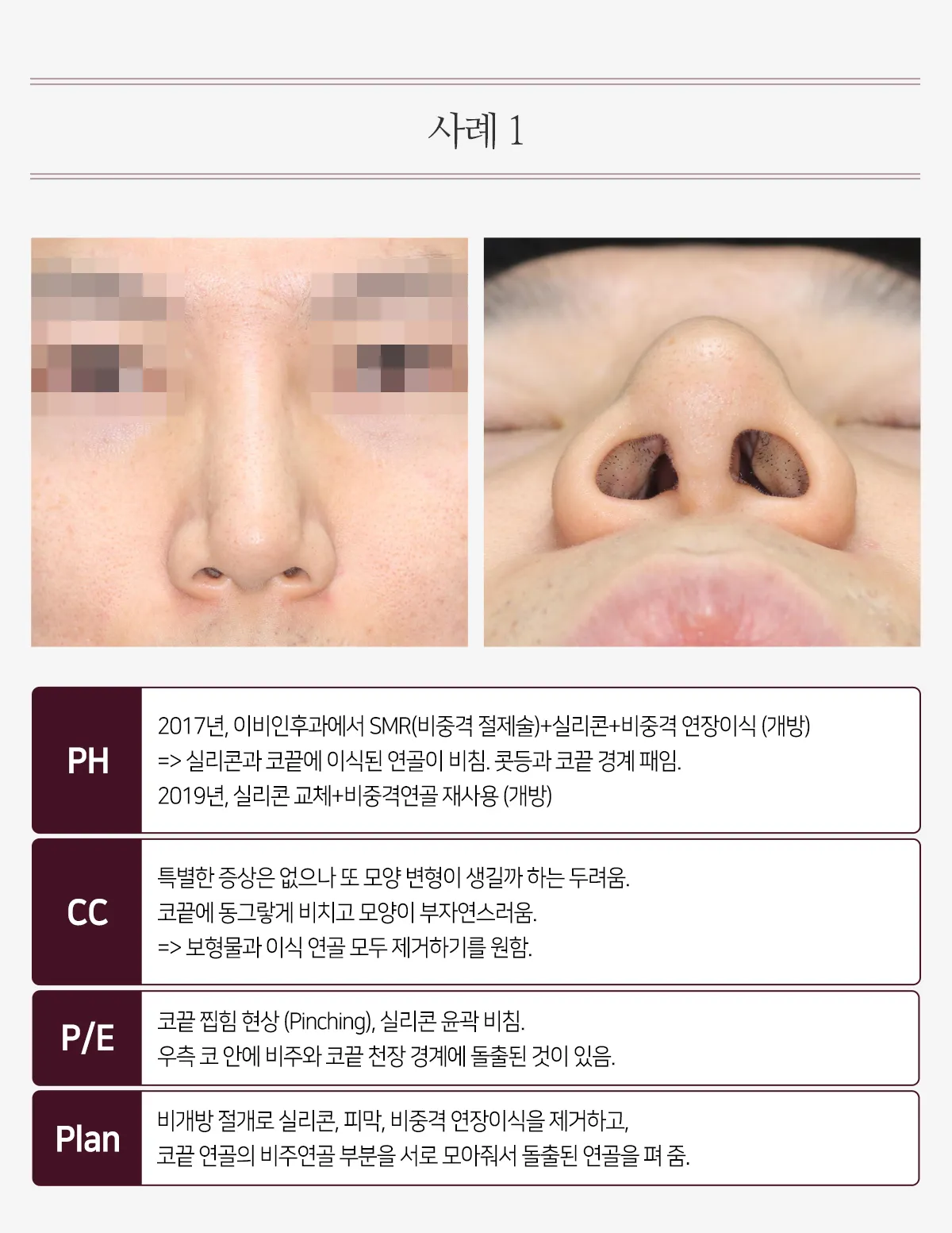
PH
2017, at an ENT clinic: SMR (septal resection) + silicone + septal extension graft (open)
=> The silicone and the cartilage grafted to the nasal tip showed through. A depression formed at the junction of the dorsum and tip.
2019: silicone replaced + septal cartilage reused (open)
CC
No particular symptoms, but fears the shape may deform again.
A round shape shows through at the nasal tip, and the shape is unnatural.
=> Wants both the implant and the grafted cartilage removed.
P/E
Pinched tip (pinching), silicone outline showing through.
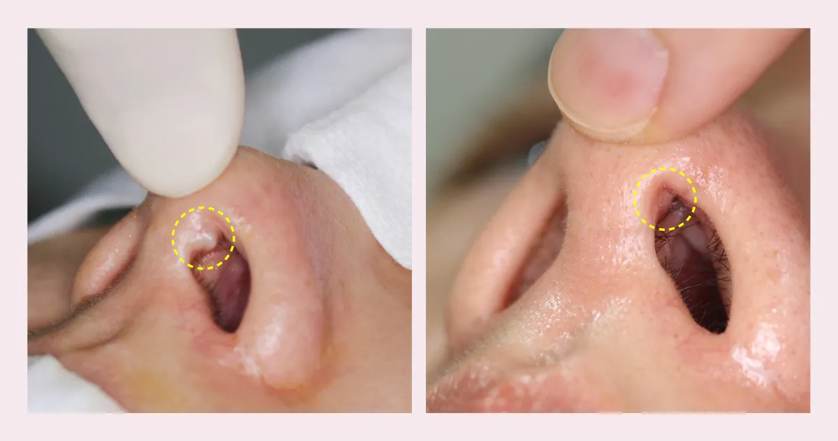
Inside the right nostril, there is a protrusion at the junction of the columella and the roof of the nasal tip.
Plan
Through a closed approach, the silicone, capsule, and septal extension graft were removed,
and the columellar cartilage portions of the tip cartilages were brought together to flatten the protruding cartilage.
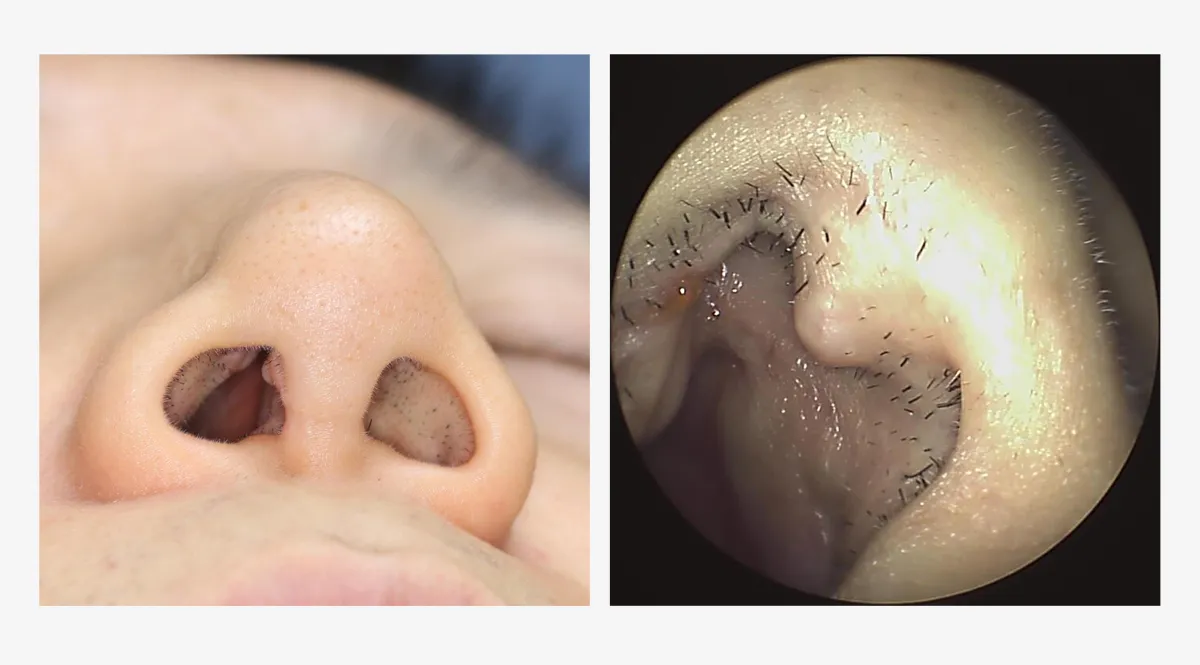
A protrusion like this can be seen inside the right nostril.
We see such patients often; they think a piece of grafted cartilage has come out,
but in most cases it is the columellar cartilage that has buckled and protrudes like this.
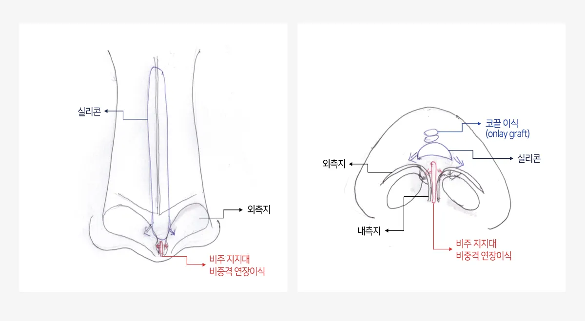
The alar cartilages of the nasal tip normally curve along the nostrils, with the lateral crus, which forms the ala,
continuous with the medial crus, which forms the columella.
However, as in the illustration, when dorsal silicone extends down to the nasal tip and presses on the tip cartilages,
the junction of the lateral and medial crura is pushed outward and protrudes inside the nose.
Another cause is a columellar strut or septal extension graft between the medial crura of the tip cartilages;
this often pushes out the area where the medial and lateral crura meet in particular, making it protrude.
The solution is to suture the buckled tip cartilages and bring them together so that they flatten out.
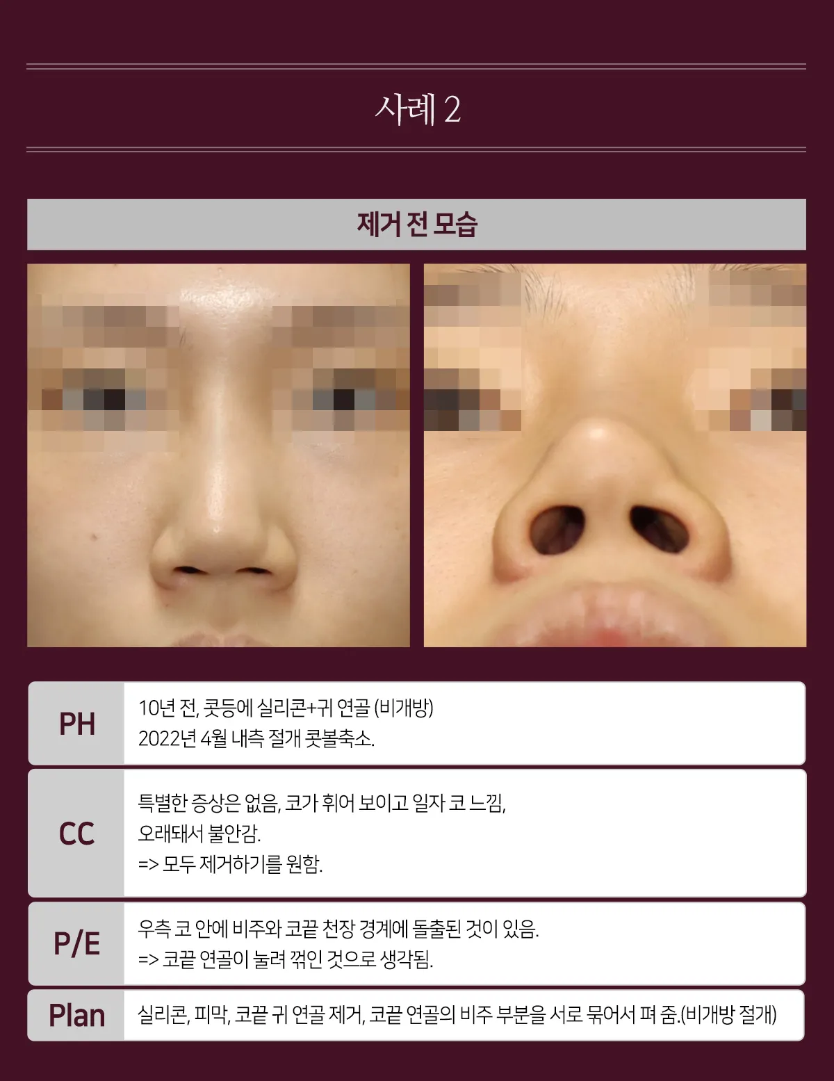
PH
10 years ago: dorsal silicone + ear cartilage (closed)
April 2022: alar reduction through an inner incision.
CC
No particular symptoms; the nose looks deviated and feels like a straight-line nose,
and anxious because it is so old.
=> Wants everything removed.
P/E
Inside the right nostril, there is a protrusion at the junction of the columella and the roof of the nasal tip.
=> Thought to be the tip cartilage pressed and buckled.
Plan
Silicone, capsule, and tip ear cartilage removed; the columellar portions of the tip cartilages sutured together and flattened. (Closed approach)
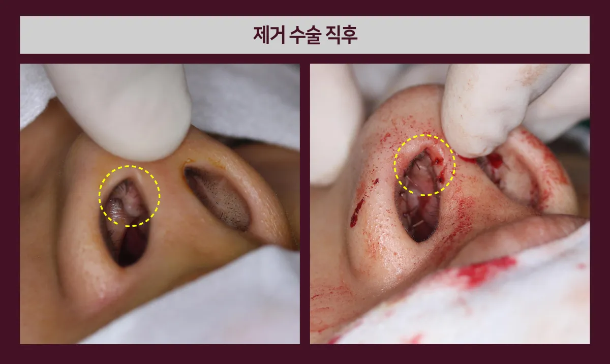
Before surgery, a protrusion is visible inside the nose at the junction of the columella and the roof of the nasal tip.
This was the tip cartilage buckled and protruding; after the silicone, capsule, and tip ear cartilage were removed through a closed approach,
the columellar portions of the tip cartilages were sutured together to straighten the buckled cartilage.
Immediately after surgery, the protrusion can be seen to be gone.
Among patients who visit us for implant removal like this,
protruding cartilage that can be felt inside the nose is often seen, and it can be corrected even with a closed approach.
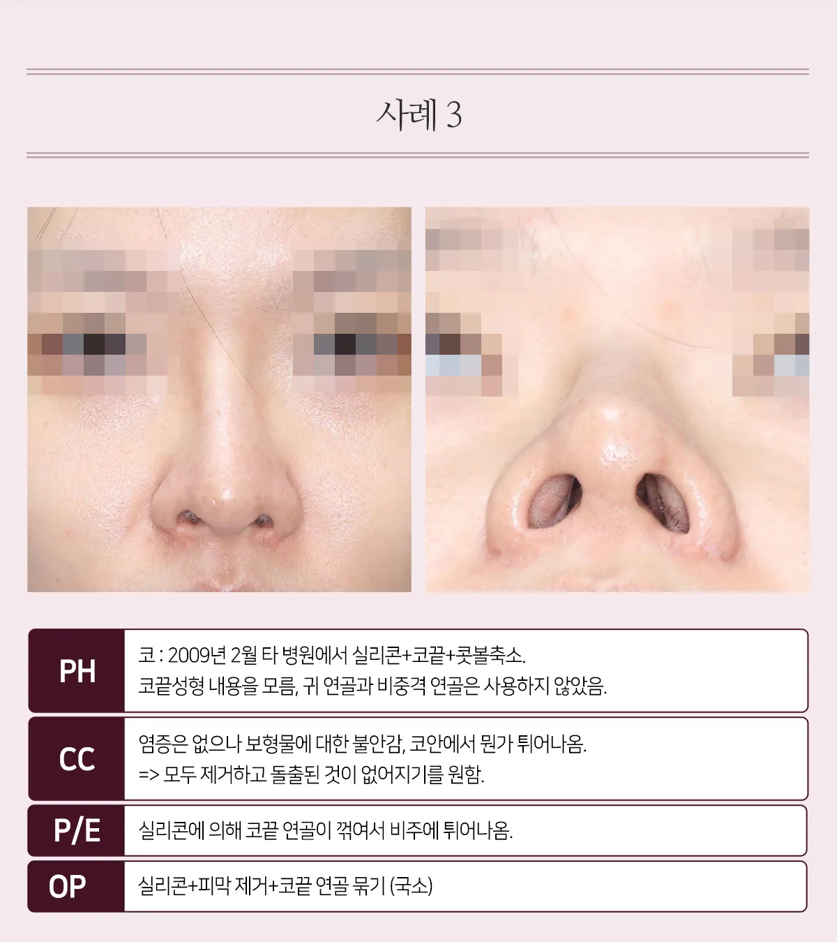
PH
Nose: February 2009 at another clinic, silicone + tip plasty + alar reduction.
Details of the tip plasty unknown; ear cartilage and septal cartilage were not used.
CC
No inflammation, but anxious about the implant; something protrudes inside the nose.
=> Wants everything removed and the protrusion gone.
P/E
The tip cartilage was buckled by the silicone and protrudes at the columella.
OP
Silicone + capsule removal + tip cartilage suturing (local anesthesia)