

Nasal Implant Extrusion and Nasal Contracture Case #ContractedNose #NasalImplantRemoval
Nasal Implant Extrusion and Contracture Case
When silicone extrudes inside the nose after rhinoplasty with silicone and autologous septal cartilage,
and there is discharge with infection, contracture develops in most cases.
In such cases, it is not easy to decide how to operate.
The following three questions are the ones most often weighed.
01
Whether removal and revision surgery (contracture correction and augmentation rhinoplasty) should be done in a single operation or divided into 2 operations
02
Whether to use an open approach or a closed approach
03
How to close the hole that has formed inside the nose
The case presented here is a patient who had augmentation rhinoplasty with silicone 8 years ago, with a septal extension graft at the nasal tip,
and who, starting 1-2 months ago, could feel the implant protruding inside the nose, with pus draining, the nose turning upward, and the contracture worsening.
PH
8 years ago: silicone augmentation rhinoplasty + tip plasty with septal cartilage + alar reduction (open approach)
▶ The nose gradually turned upward.
1 month ago, after a blow to the nose, the nasal tip became red and swollen, and pus began to drain from inside the nose.
P/E
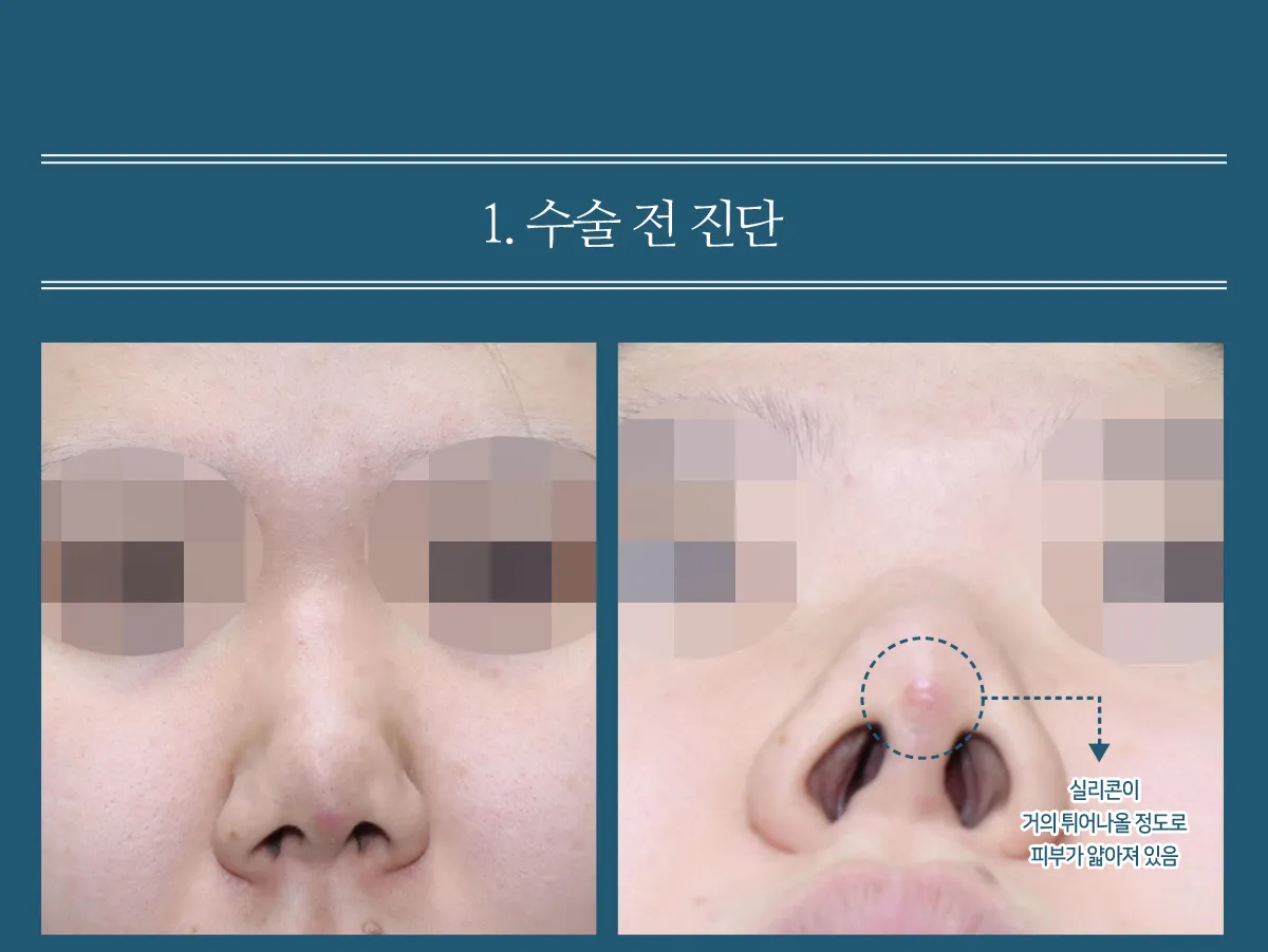
Nostril show.
The nasal tip was upturned and deformed.
The skin over the upper part of the columella had thinned almost to the point of perforation, and the implant showed through.
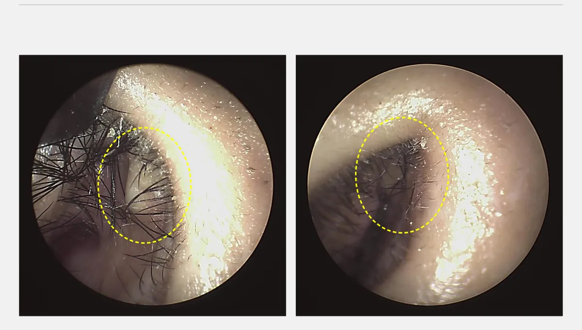
A hole had formed in the mucosa inside the left side of the nose, exposing the silicone. (Endoscopic photo)
The dorsum was thick and bunched up because of contracture and scar tissue.
OP
Through a closed approach, the silicone, capsule, scar tissue, and inflamed tissue were removed; after tip cartilage repositioning, the hole in the mucosa was sutured.
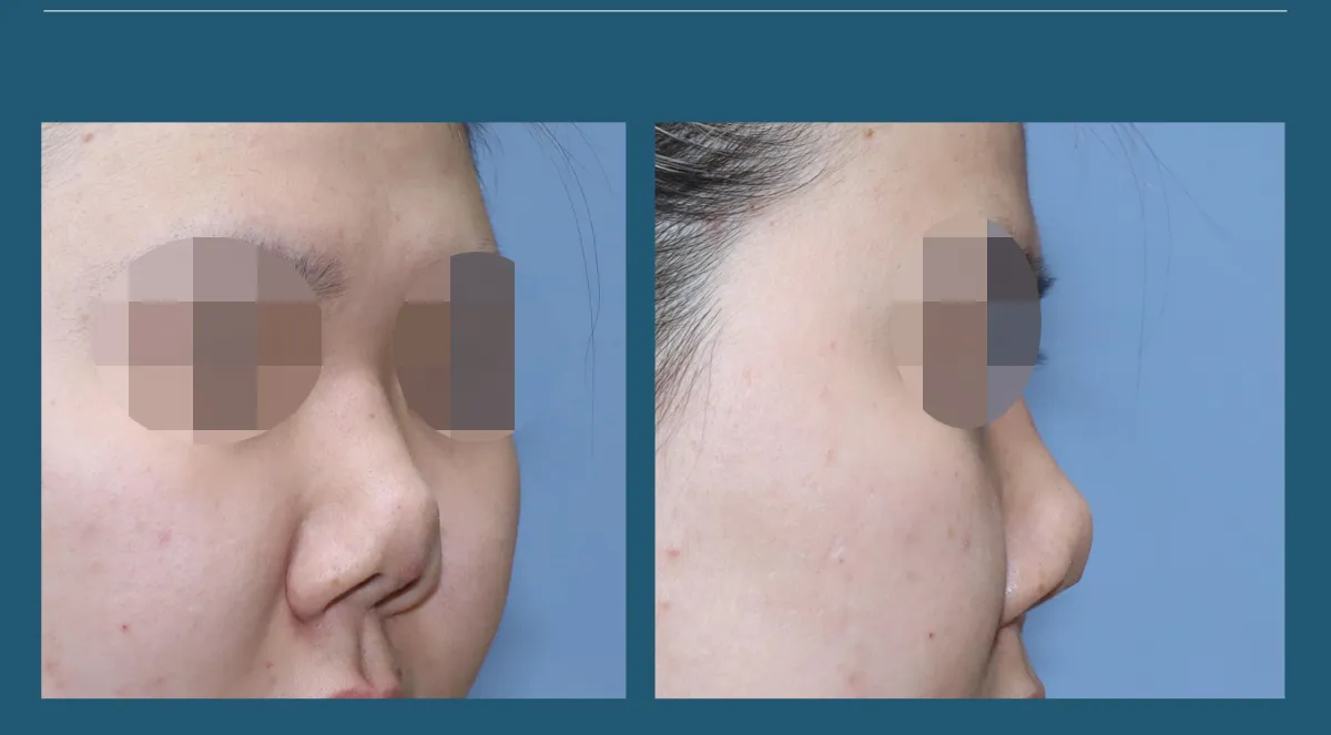
The nasal tip looks cut off, as if severed, because of contracture, and is markedly upturned.
The soft tissue of the dorsum is thick because inflamed tissue and scar tissue have clumped together.

Inside the left side of the nose, the silicone implant can be seen breaking through at the upper part of the columella.
The hole seen on endoscopy was about 7 mm in diameter, and on the columella inside the right side of the nose, the cartilage can be seen bent and protruding.
Preop
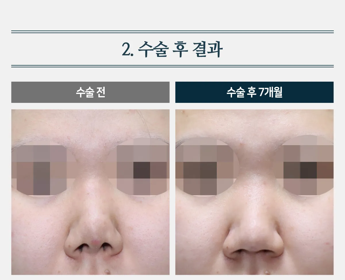
The nasal tip was upturned and uneven, with nostril show.
Because of infection and contracture, the dorsum was uneven, thick, and blunt.
Postop
The upturned tip and the irregularities were resolved.
The nostril show is gone.
The dorsum became slim and straight.
OP
Through a closed approach, the silicone, capsule, scar tissue, and inflamed tissue were removed; after tip cartilage repositioning, the hole in the mucosa was sutured from inside the nose.
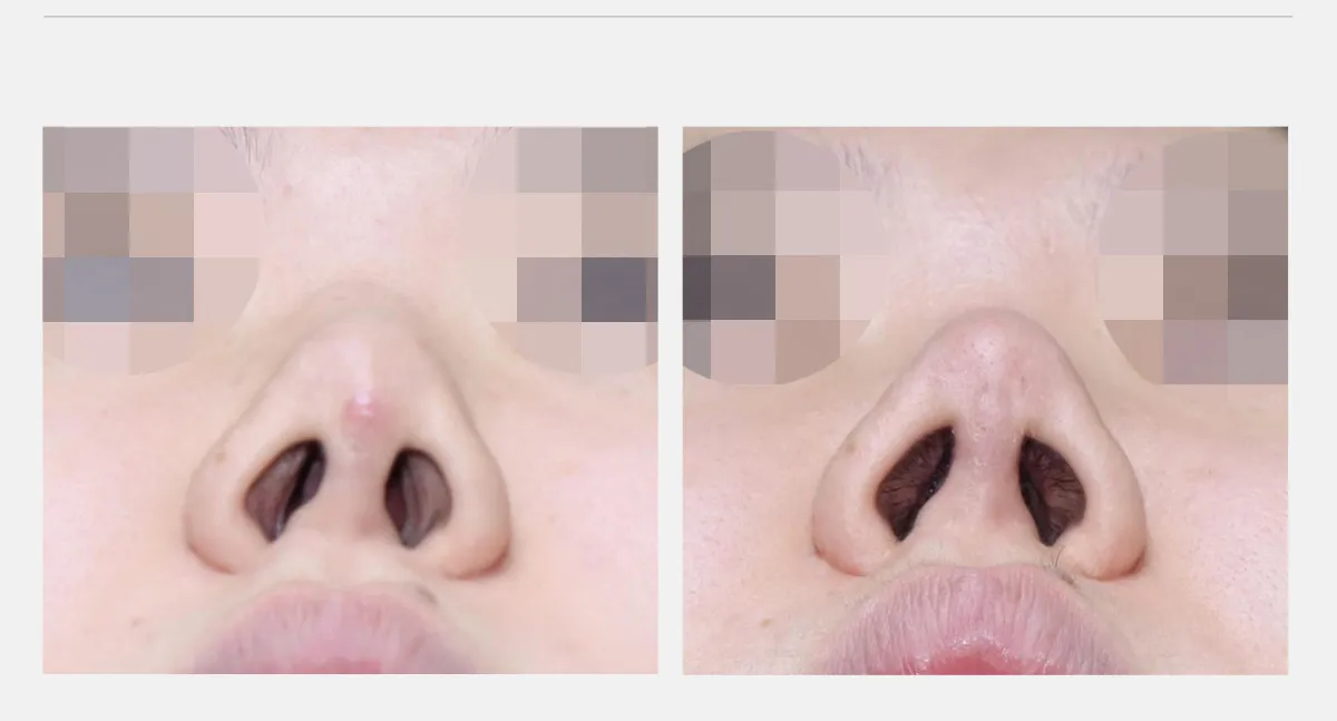
The skin of the nasal tip had thinned almost to the point of perforation and was red with an inflammatory response.
After the removal surgery, the nasal tip healed well without scarring, and the severely deformed tip shape has returned to a natural appearance.
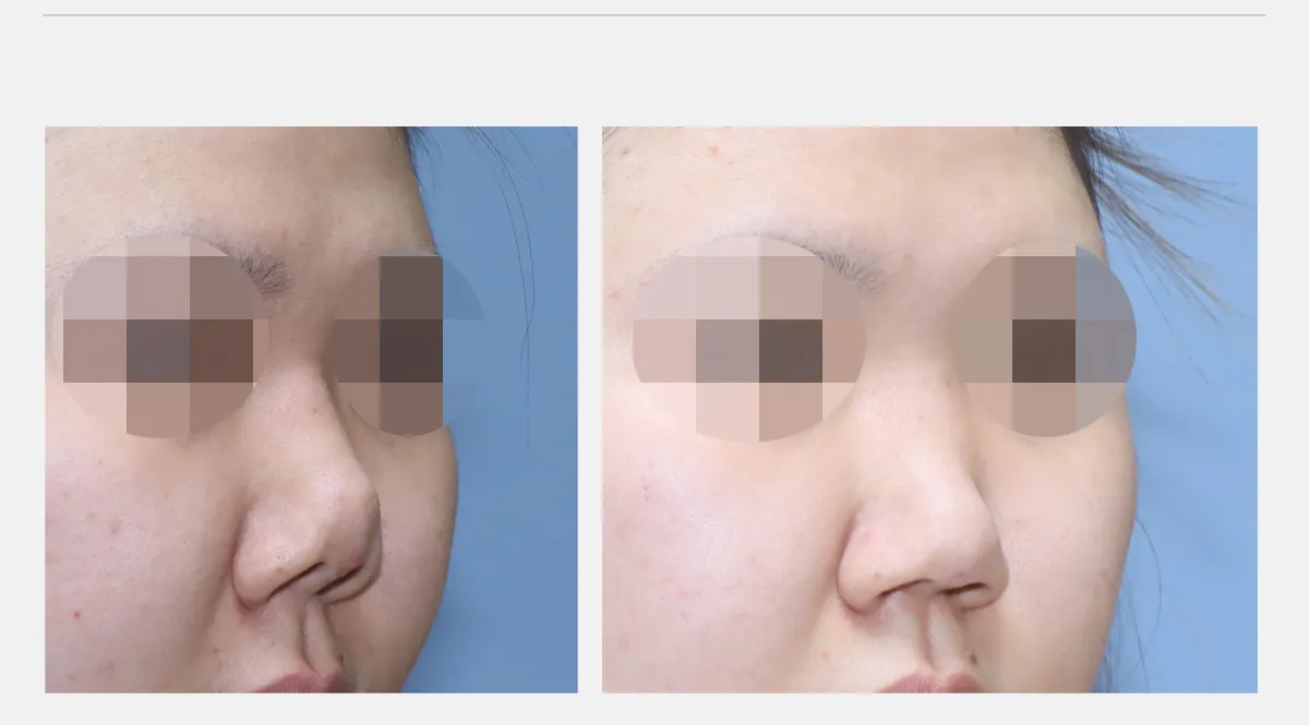
The contour that appeared to break off between the dorsum and the tip became natural after removal and repositioning.
By removing the silicone, the capsule, and the surrounding scar tissue to release the contracture, the length of the nasal tip was naturally restored.
The uneven dorsum has also become slimmer.
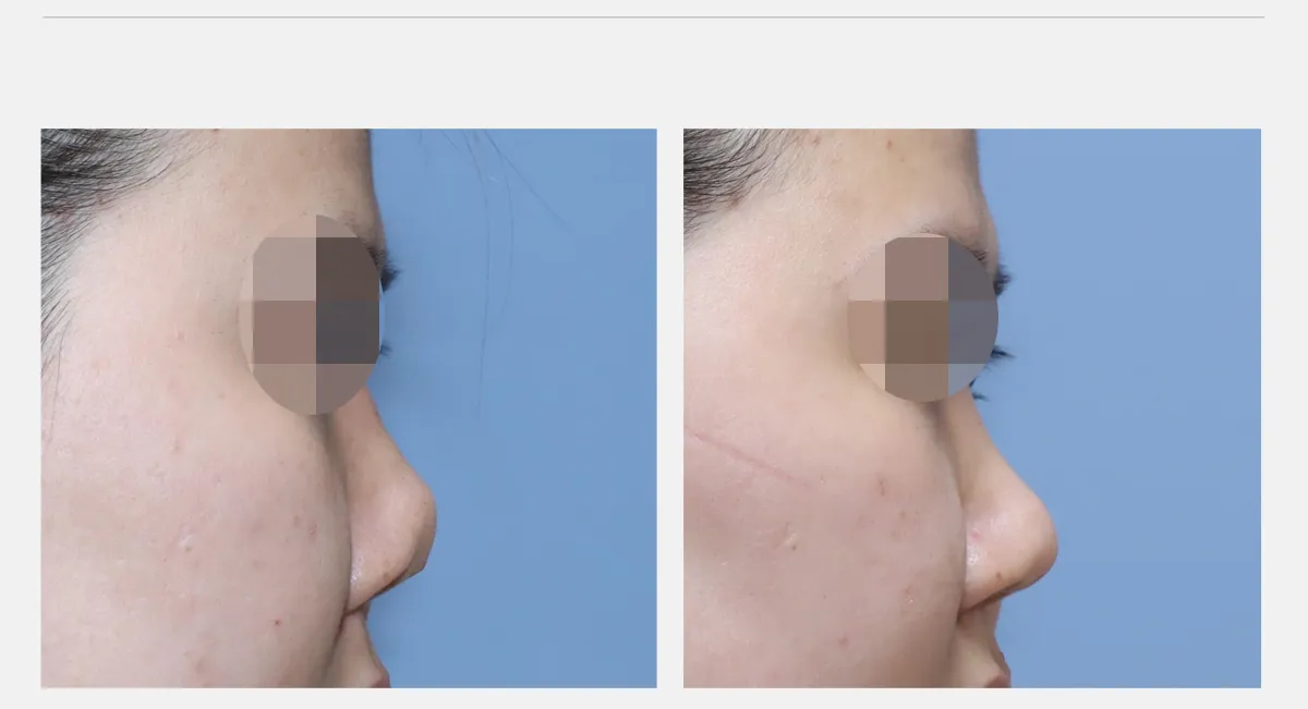
The tip, which had looked cut off, has naturally regained its length and shape.
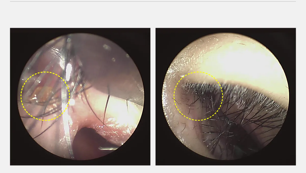
The area where a hole had formed and the silicone implant was visible has healed cleanly.
The columellar cartilage at the tip, which had been bent under pressure from the implant, has straightened after repositioning.