

Revision Rhinoplasty Cases by Type (Contracted Nose Caused by Silicone and Inflammatory Response) 1
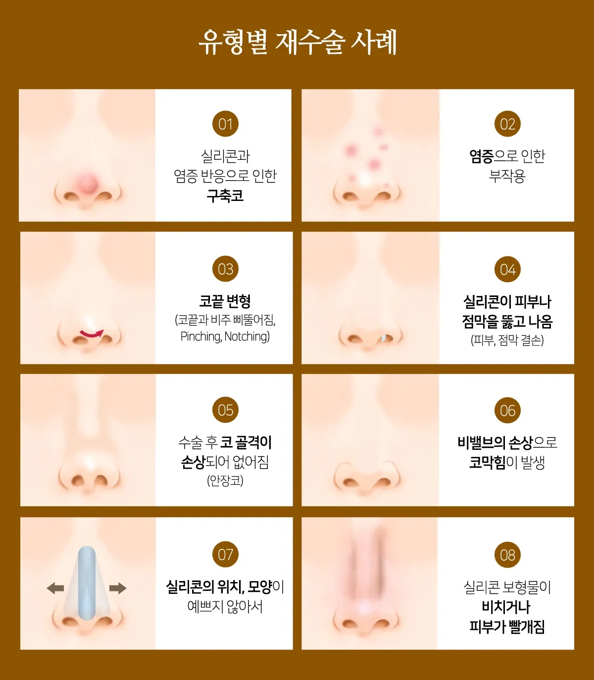
Revision rhinoplasty refers to corrective surgery performed when, after one or more nasal surgeries,
aesthetic or functional side effects have developed.
The deformities that result from problems after rhinoplasty are so varied
that it is difficult to show every case one by one.
The reason is that the nose is a three-dimensional structure and a complex organ in which various tissues are organically connected.
We will therefore focus on representative cases and explain how, after revision surgery, side effects can be kept from recurring
and a good result can be achieved.

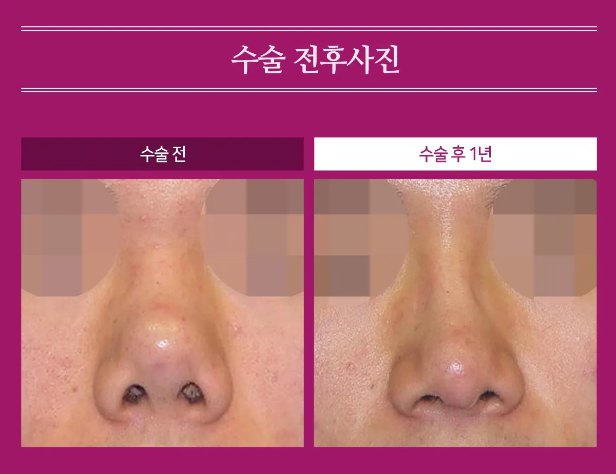
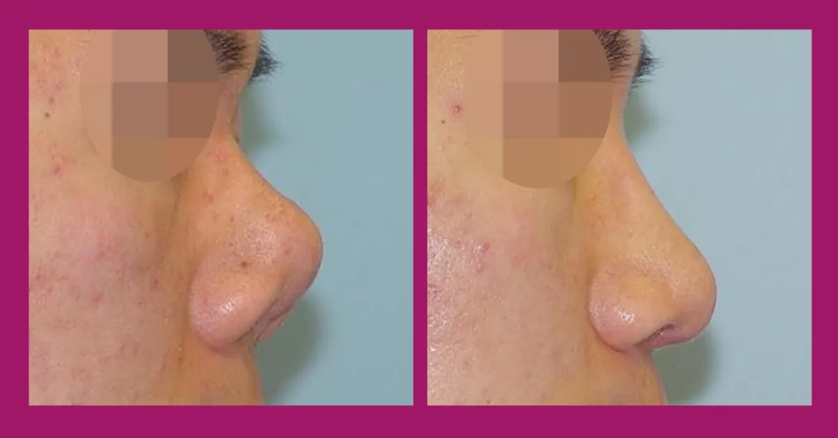
After removing the scar tissue and performing wide dissection to release the shrunken skin and framework of the tip,
the nose was lengthened using autologous costal cartilage.
The dorsum was also augmented with costal cartilage, and the nasal length, dorsal height, and other aspects all improved to a satisfactory degree.
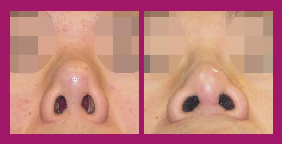
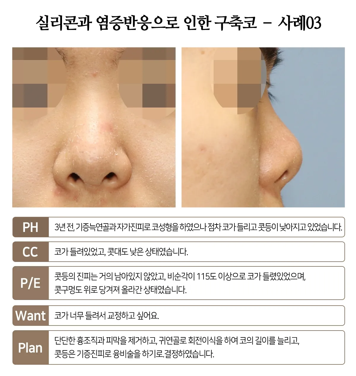
Before surgery, the tip was pulled upward by the contracture,
but in the photo taken after the contracture was corrected, the tip is naturally relaxed.

The left-side view after surgery shows sufficient length and a natural bridge.

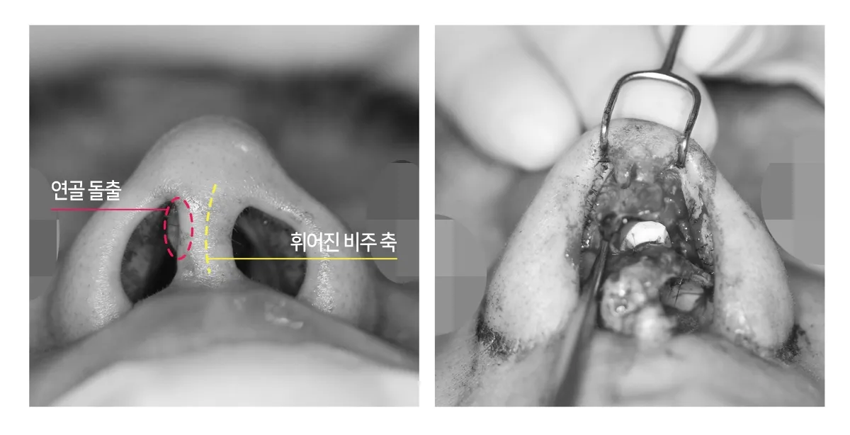
Before surgery, the columella was crooked, something could be seen protruding inside the right nostril,
and there was slight pinching and notching.
The right photo, taken while the silicone was being removed, shows that the tip cartilages were twisted.
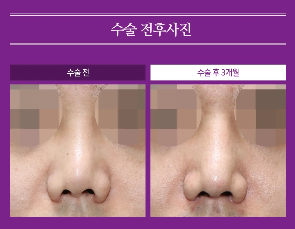
The dorsum, which was crooked before surgery, has been straightened, and the nose has been lengthened.

Before surgery, the columella was crooked, which made the nostrils asymmetric,
but after surgery the columella was straightened, and it and the nostrils all became symmetric.

The side view confirms that the contracture has improved; the nose has been sufficiently lengthened,
and the dorsum now has a natural, masculine appearance.
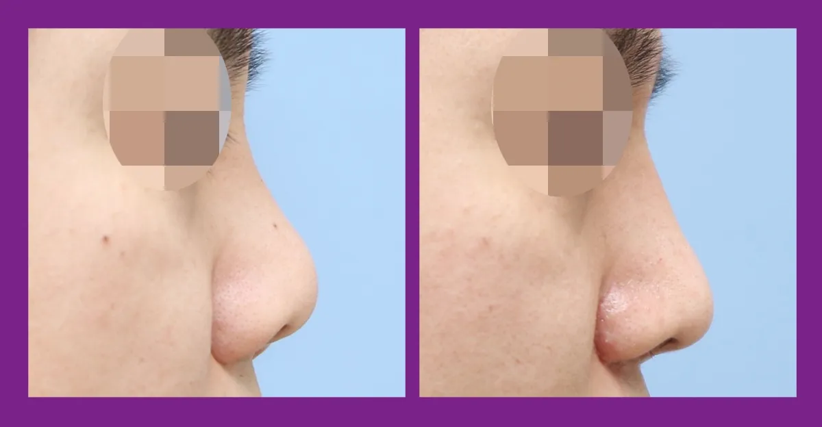
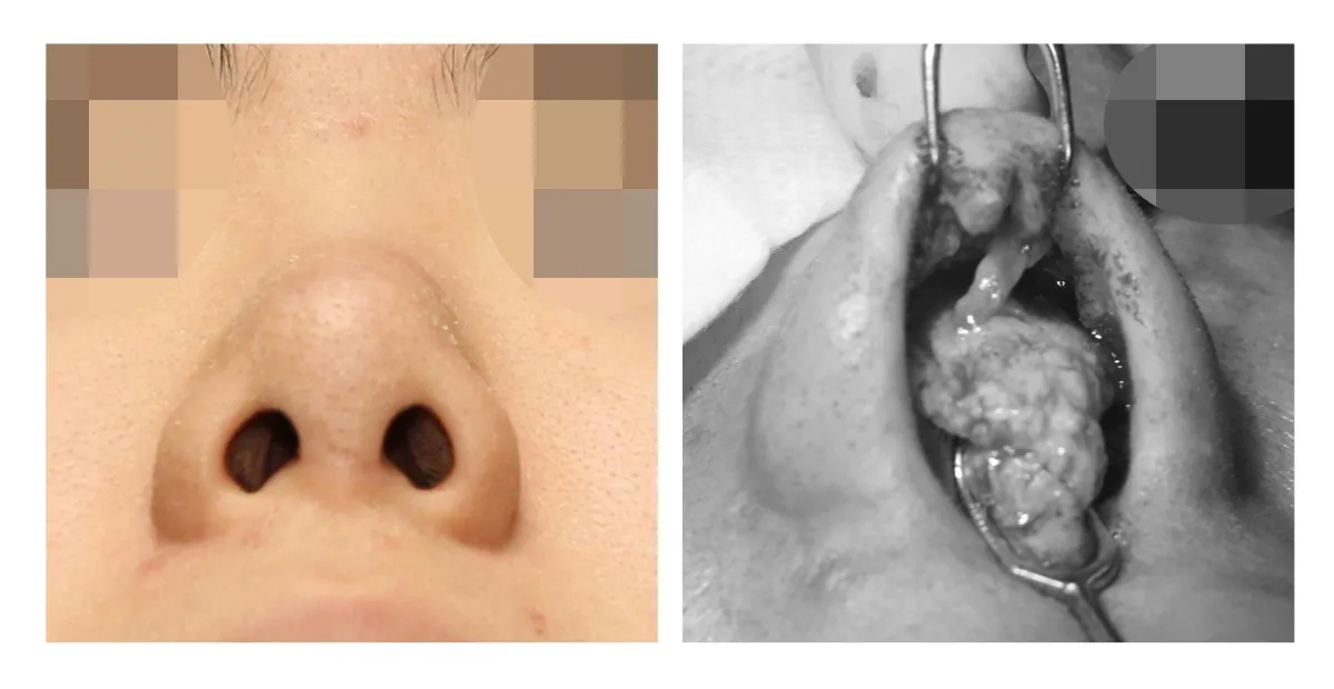
Before surgery, the columella was severely bent, and there was a good deal of thickened tissue at the tip.
After the dermal tissue that had caused the contracture and the thickened tissue were removed to fully release the tip, a derotation graft was placed using ear cartilage.

The dorsum was given natural height using acellular dermal matrix.
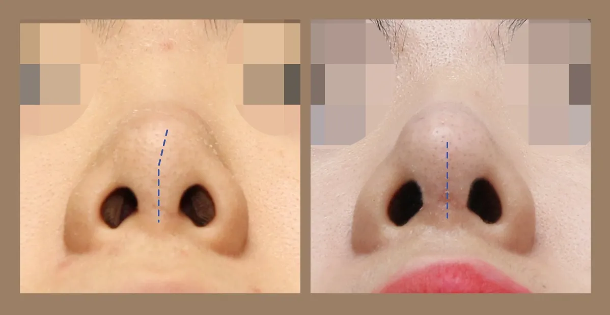
The blunt, heavy tip has become well defined, and the tilt of the tip and columella has also improved.
The nostrils have naturally become symmetric.
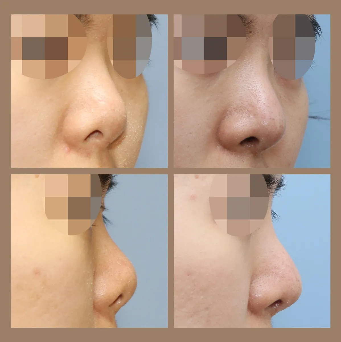
The upturned tip has come down so that the nose is sufficiently lengthened, and the bridge has been raised to an appropriate height.