

Resolving Entropion Caused by Repeated Lateral Canthoplasty

After lateral canthoplasty, some patients are not satisfied with how much larger the eyes became, the opened area closes up again,
or the opening is excessive, and so the lateral canthoplasty is repeated several times.
When lateral canthoplasty, which opens the outer corner to lengthen and widen the eye, is repeated in this way,
the tissue of the tarsal plate is damaged, the outer corner gapes and sags,
and entropion develops, in which the tarsal plate rolls in toward the eyeball.
Resolving this is quite demanding and requires a high level of skill and concentration.
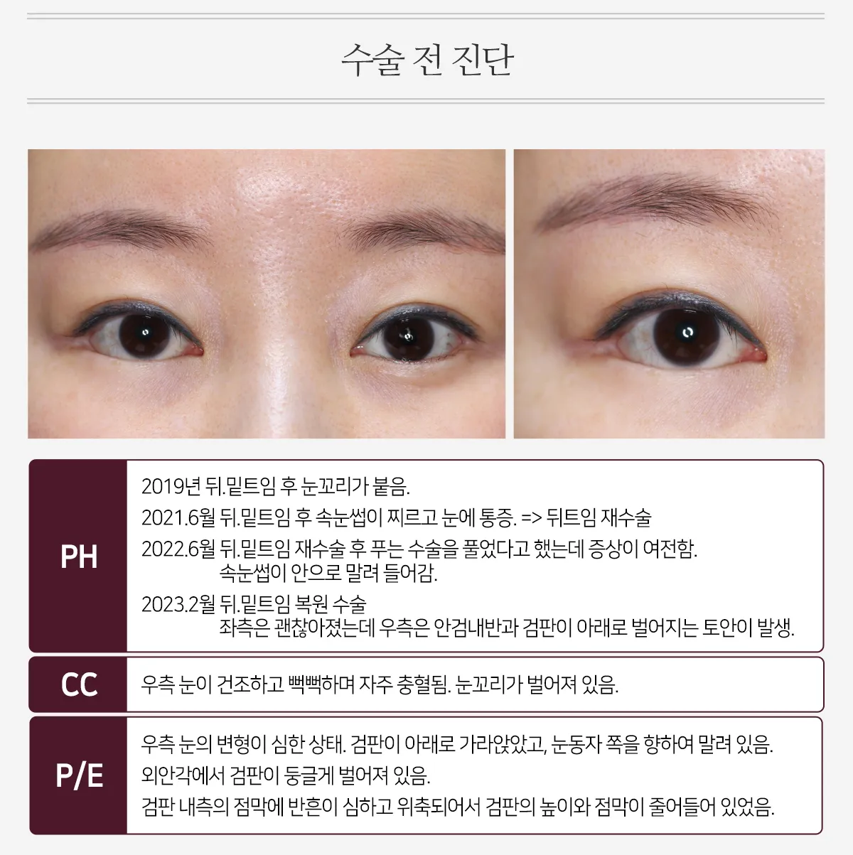
PH
2019: After lateral and lower canthoplasty, the outer corners closed up again.
June 2021: After lateral and lower canthoplasty, the eyelashes poked the eye and caused pain. => Revision lateral canthoplasty
June 2022: After revision lateral and lower canthoplasty, the patient was told that a release procedure had been done, but the symptoms persisted. The eyelashes turned inward.
February 2023: Lateral and lower canthoplasty reversal
The left side improved, but the right side developed entropion and lagophthalmos, with the tarsal plate pulled open downward.
CC
The right eye was dry and gritty and often became red. The outer corner was gaping.
P/E
The right eye was severely deformed. The tarsal plate had sunk downward and was rolled in toward the eyeball.
At the lateral canthus, the tarsal plate was splayed open in a rounded shape.
The mucosa on the inner side of the tarsal plate was heavily scarred and atrophied, so both the height of the tarsal plate and the amount of mucosa were reduced.
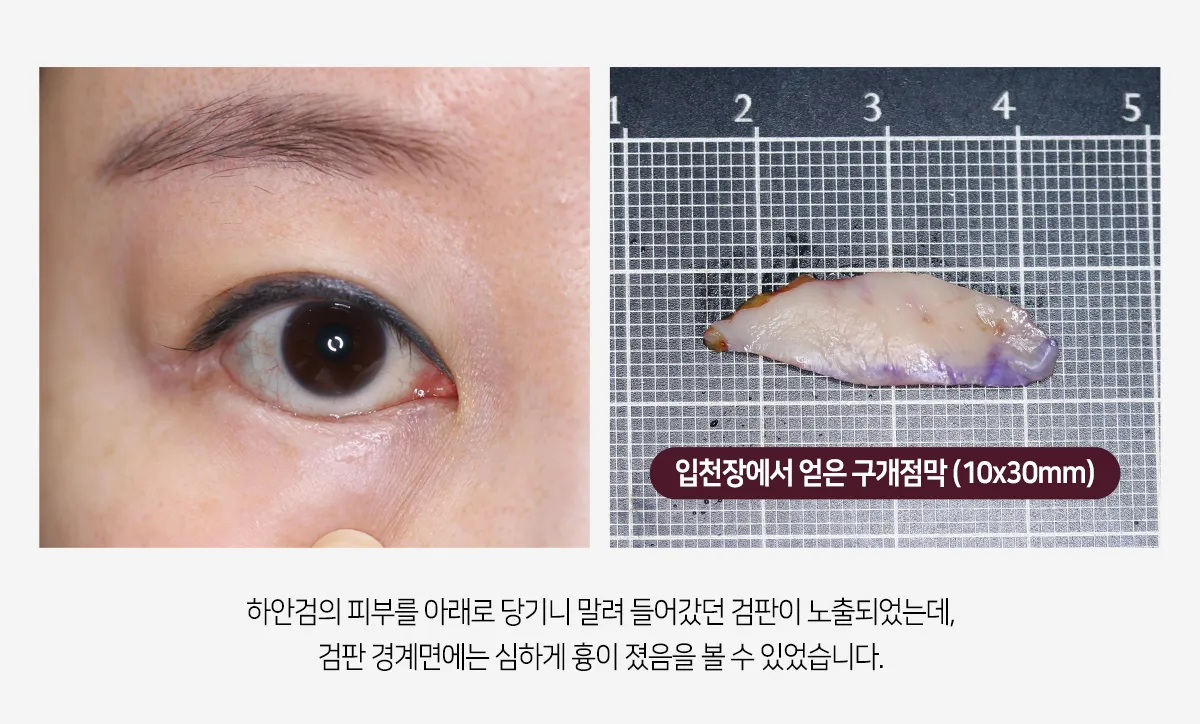
When the lower eyelid skin was pulled downward, the tarsal plate that had rolled inward was exposed,
and severe scarring could be seen along its margin.
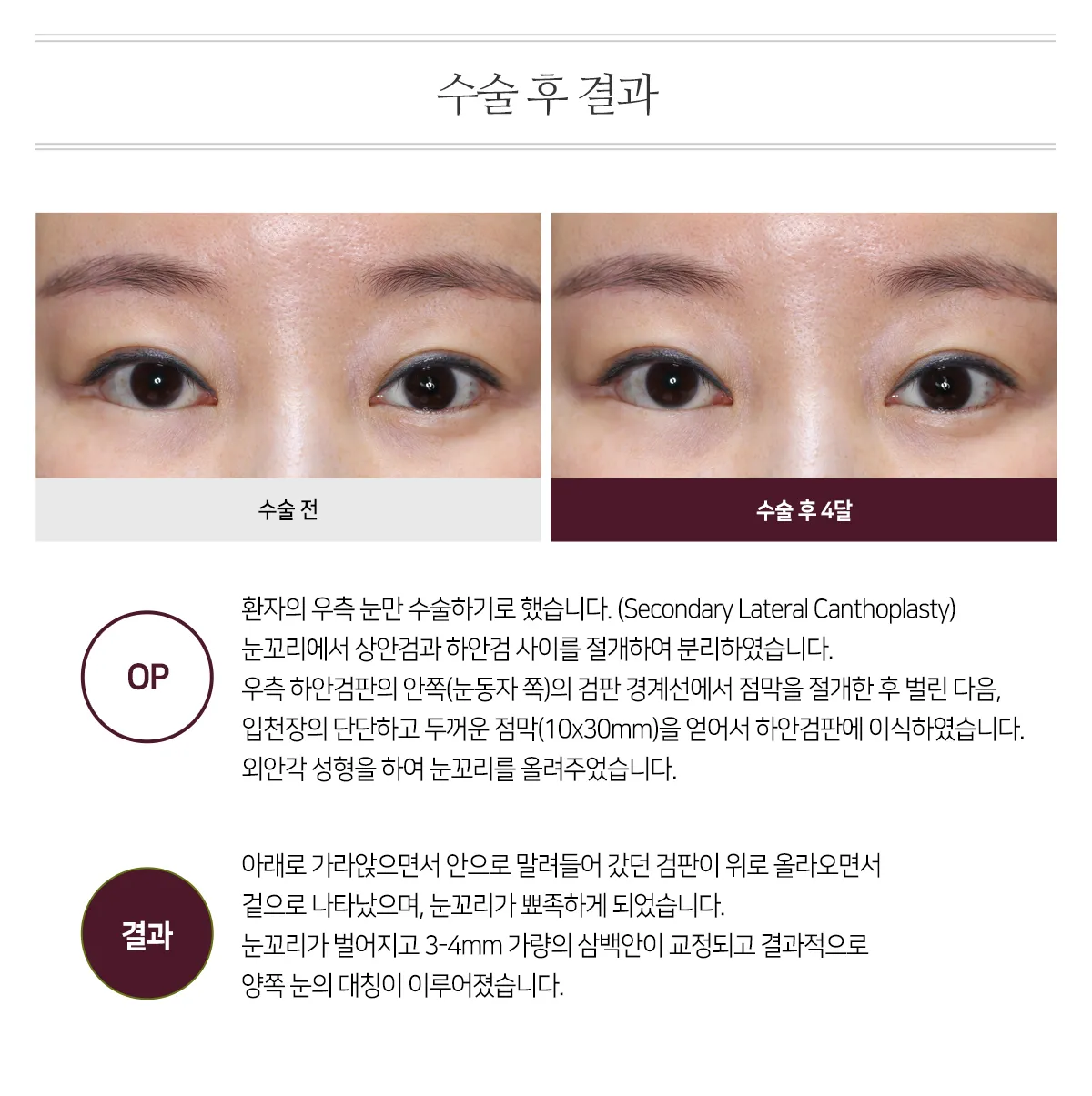
OP
We decided to operate only on the patient's right eye. (Secondary Lateral Canthoplasty)
At the outer corner, an incision was made between the upper and lower eyelids to separate them.
On the inner (eyeball) side of the right lower tarsal plate, the mucosa was incised along the tarsal border and spread open,
and firm, thick mucosa from the palate (10x30mm) was harvested and grafted onto the lower tarsal plate.
Lateral canthal reconstruction was then performed to raise the outer corner.
Results
The tarsal plate, which had sunk downward and rolled inward, moved back up
and became visible again, and the outer corner took on a pointed shape.
The gaping outer corner and about 3-4mm of sanpaku eye were corrected, and as a result
the two eyes became symmetrical.
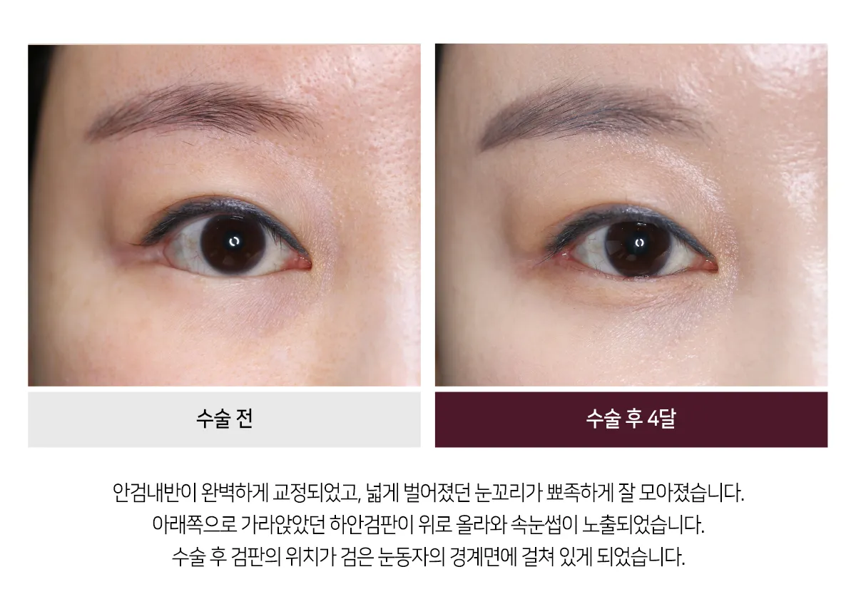
The entropion was completely corrected, and the widely gaping outer corner came together neatly into a point.
The lower tarsal plate, which had sunk downward, moved up, and the eyelashes became visible.
After surgery, the tarsal plate came to rest at the edge of the iris.
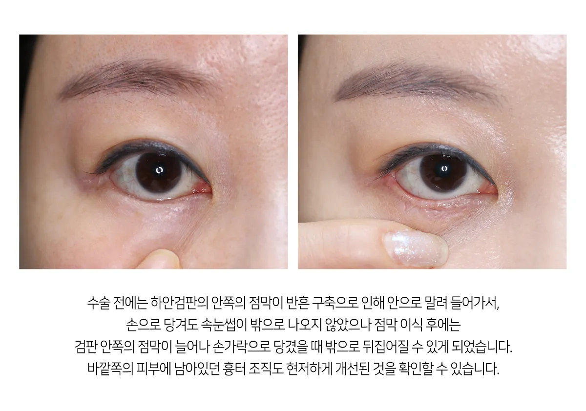
Before surgery, the mucosa on the inner side of the lower tarsal plate was rolled inward by scar contracture,
so the eyelashes did not come out even when the lid was pulled by hand; after the mucosal graft, however,
the mucosa on the inner side of the tarsal plate was lengthened, and the lid could be everted when pulled with a finger.
The scar tissue that had remained on the outer skin can also be seen to have improved markedly.
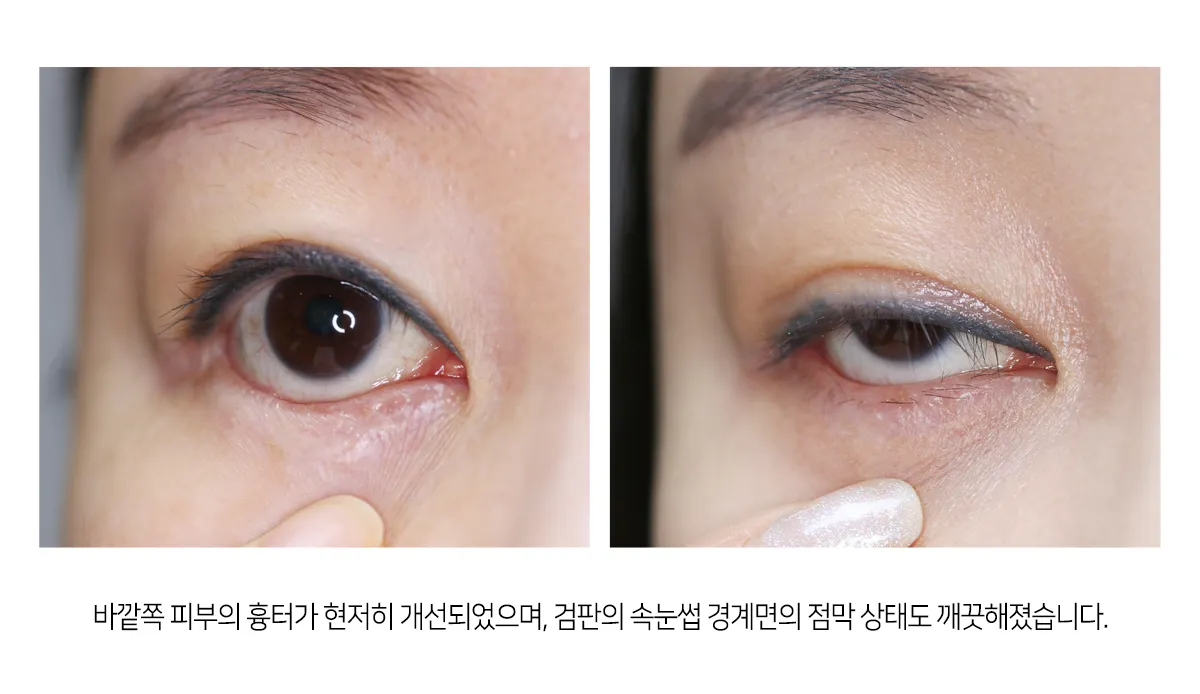
The scar on the outer skin improved markedly, and the mucosa along the lash margin of the tarsal plate also became clean.
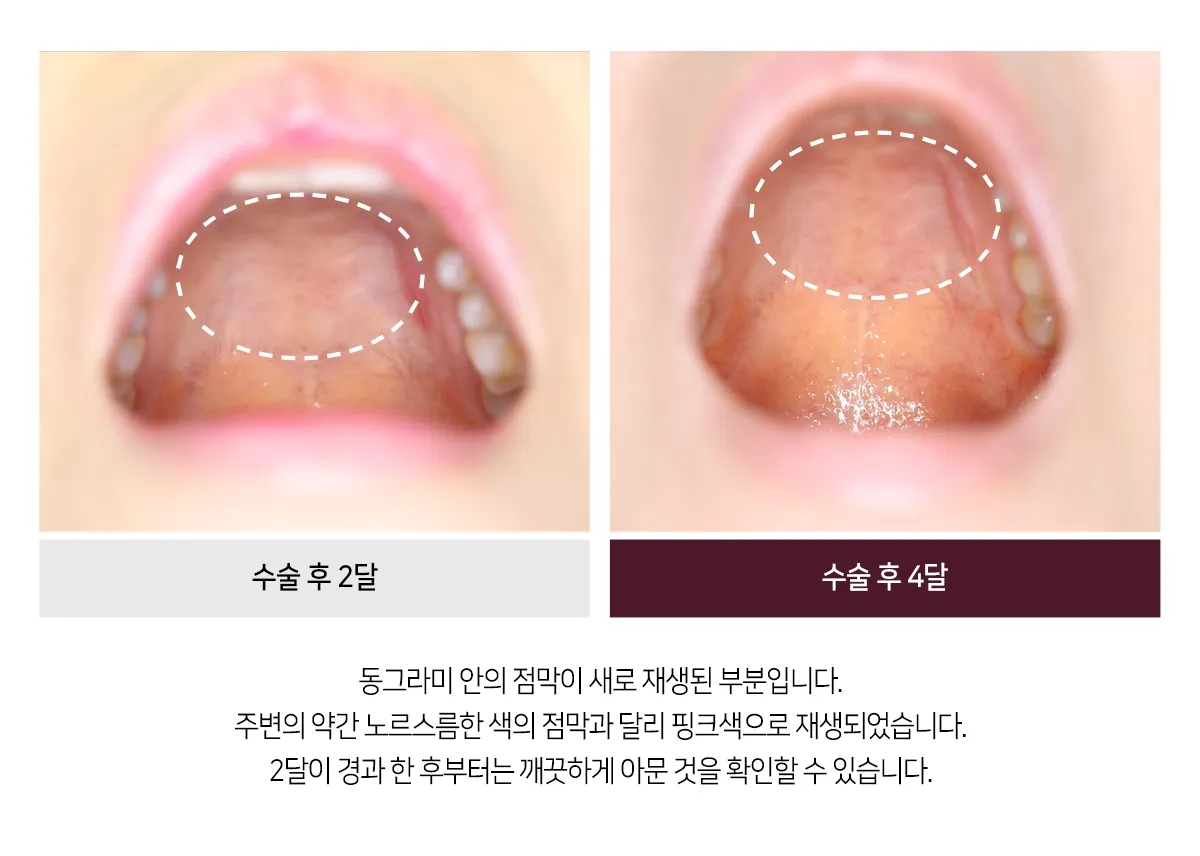
The mucosa inside the circle is the newly regenerated area.
Unlike the slightly yellowish surrounding mucosa, it regenerated in a pink color.
From 2 months after surgery, it can be seen to have healed cleanly.