

Dorsal Hump Case Corrected by Silicone Removal and Revision Surgery

Resembling a hawk's beak,
with a prominently raised dorsum, the"dorsal hump"
is a source of self-consciousness about appearance for some people,
and many undergo rhinoplasty
to correct it.
However, after dorsal hump correction,
some are left with unsatisfactory results
and wish to undergo revision rhinoplasty.
to correct it.
Today we present one such case.

PH :
1 year ago, at another clinic,
the patient had augmentation rhinoplasty with "silicone" & rasping of the dorsal hump (rasp),
and, after septal resection,
an extension graft with septal cartilage and donor costal cartilage,
and a tip cap graft with ear cartilage.
After the septum was splinted with silicone sheets for 2 weeks,
cartilage became exposed through the intranasal mucosa.
P/E :
Because of a defect in the left septal mucosa,
the donor costal cartilage was exposed.
OP :
Removal of the silicone implant, capsule, and scar tissue
+ removal of 3 layers of ear cartilage at the tip
+ removal of the septal extension graft (donor costal cartilage, septal cartilage)
Temporalis fascia graft to the Lt mucosal defect
Suturing of the Rt mucosal laceration
Septal cartilage reinforcement (reusing the septal cartilage removed from the Rt side)
Columellar reinforcement with septal cartilage
For the side effects that arose from the dorsal hump correction,
surgery was planned as outlined above.

This shows the deep temporal fascia being harvested;
ear cartilage was wrapped in the fascia
to make a sandwich-like graft,
placed into the septal defect,
and sutured in place.

With ear cartilage and temporalis fascia,
the septal defect is closed.
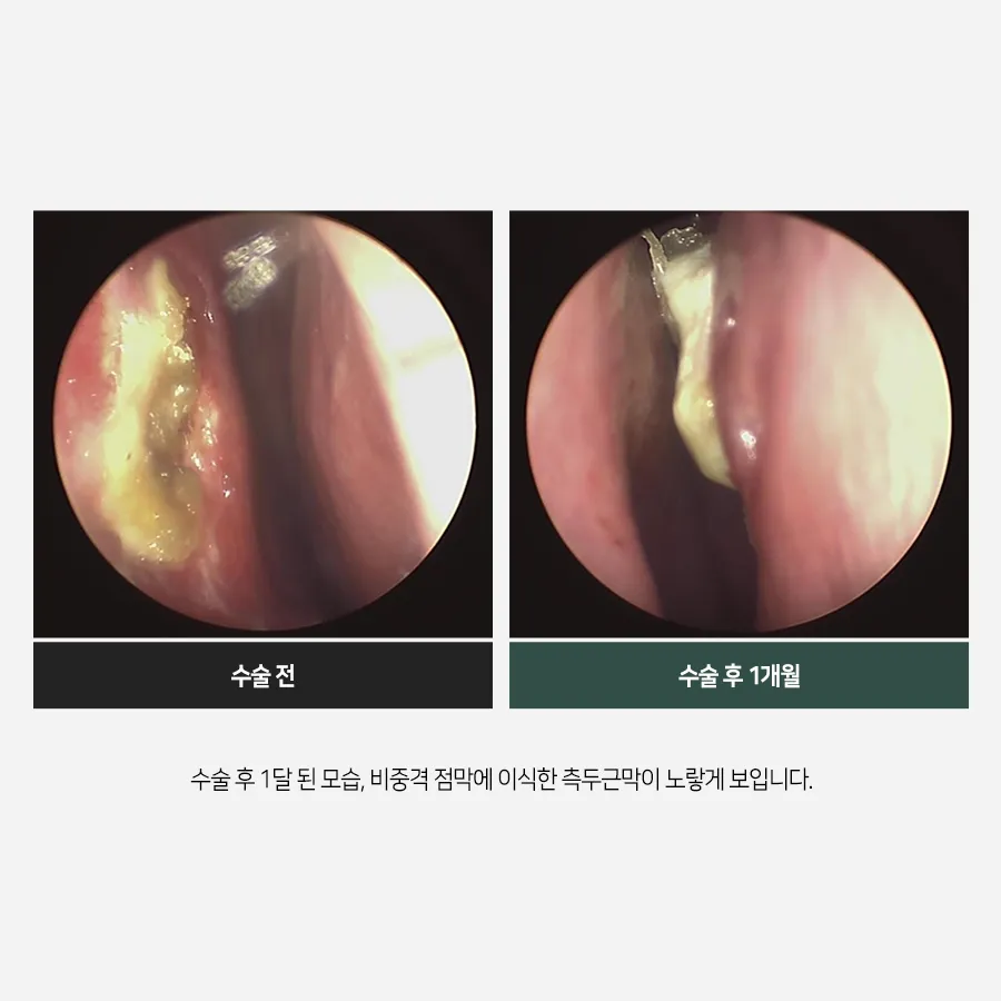
This is one month after surgery.
The temporalis fascia grafted to the septal mucosa
appears yellow.


At 4 months,
the grafted fascia looks whitish,
but it has adhered well to the mucosa and is surviving.
===============================================================
This case shows that the septal mucosal defect
was repaired successfully.