Correction of Various Nasal Tip Deformities #6 External Valve Insufficiency #Revision Rhinoplasty

External Valve Insufficiency
A distinctive feature of our clinic is that many patients, after a first surgery at another clinic went wrong,
come to us for revision surgery.
Patients therefore come with a wide range of problems,
Among these, “nasal tip deformity” is, even within the field of rhinoplasty,
the most demanding area, and one in which good results are not easy to achieve.
The reason is that “nasal tip deformity”
i) takes many different forms and is complex because the deformity appears in 3 dimensions.
ii) the way it develops and its causes are also complex, including “contracture,” “damage and deformity of the tip cartilages from repeated surgery,” and “inflammation”
among others, which makes the surgery very demanding and difficult.
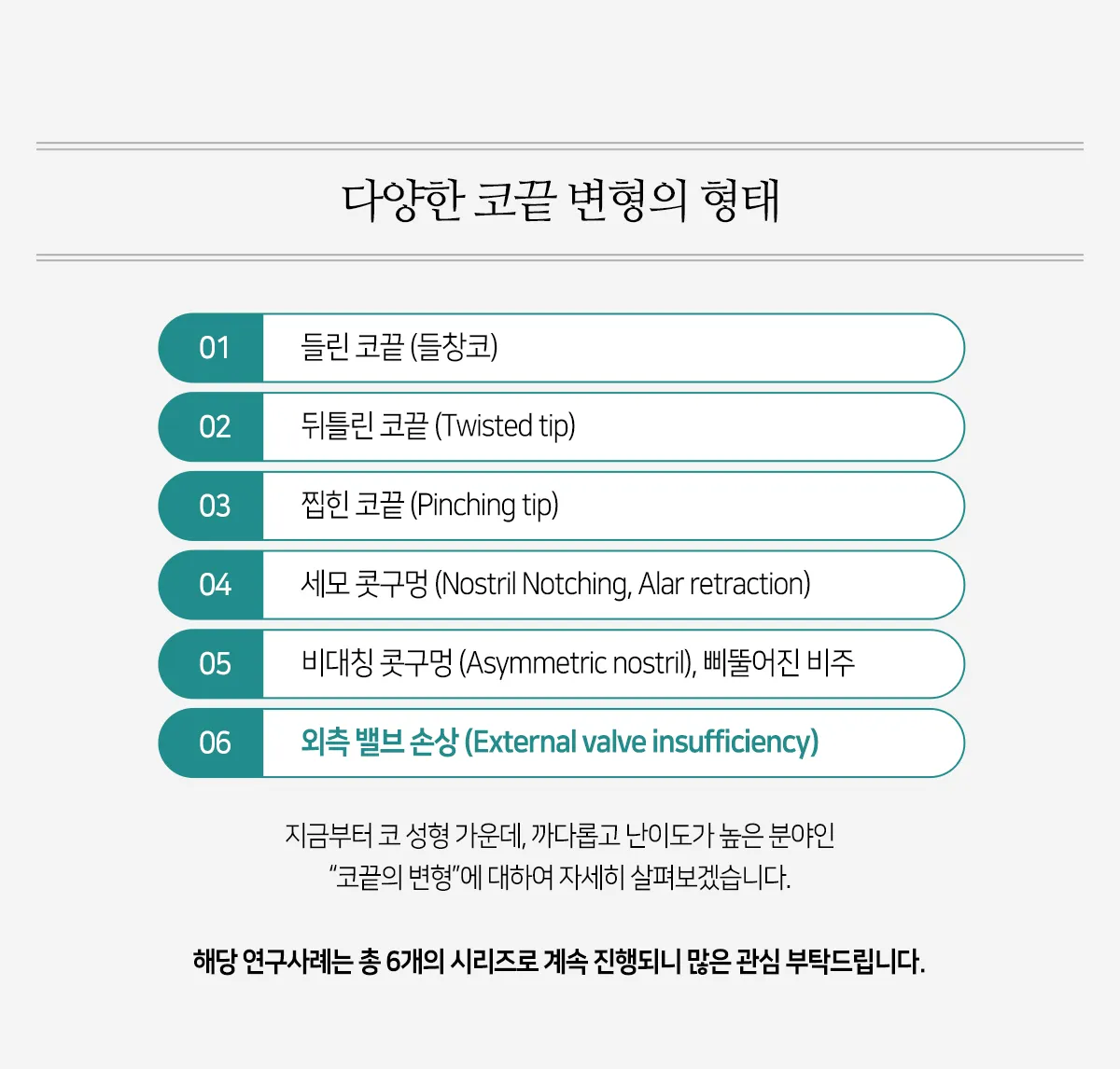
01 Upturned Tip (Upturned Nose)
02 Twisted Tip
03 Pinched Tip
04 Triangular Nostril (Nostril Notching, Alar Retraction)
05 Asymmetric Nostrils, Deviated Columella
06 External Valve Insufficiency
From here, we turn to a demanding and highly difficult area of rhinoplasty,
“nasal tip deformity,” and examine it in detail.
This case study continues as a series of 6 parts in total; we hope you will follow along.
 The external valve is the area where the tip cartilages meet the dorsal cartilages.
Because it is the junction between the two cartilages, this area weakens easily. The external valve is the area where the tip cartilages meet the dorsal cartilages. Because it is the junction between the two cartilages, this area weakens easily. When this area buckles inward, the region near the tip cartilages looks like a roof that has caved in, and because both alae are drawn in as if hollowed when breathing in, nasal breathing becomes difficult.
When this area buckles inward, the region near the tip cartilages looks like a roof that has caved in,
and because both alae are drawn in as if hollowed when breathing in, nasal breathing becomes difficult.
The external valve is the area where the tip cartilages meet the dorsal cartilages.
Because it is the junction between the two cartilages, this area weakens easily.
When this area buckles inward, the region near the tip cartilages looks like a roof that has caved in,
and because both alae are drawn in as if hollowed when breathing in, nasal breathing becomes difficult.
(Causes)
In many cases, the tip cartilages are bent excessively when they are sutured together,
or the alar framework weakens as the tip cartilages are drawn toward the center.
(Solutions)
Lateral crural repositioning, lateral crural strut graft, external valve graft
Septal cartilage / costal cartilage / ear cartilage
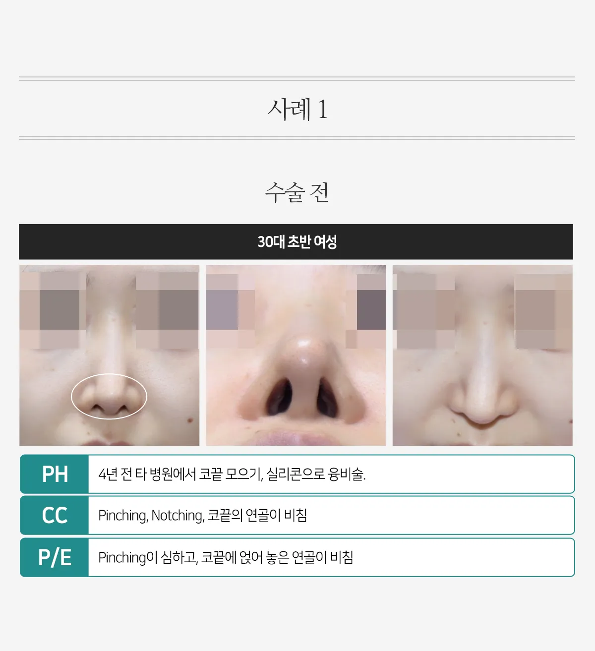
PH
4 years earlier, the patient underwent tip narrowing and augmentation rhinoplasty with silicone at another clinic.
CC
Pinching, notching, and cartilage showing through at the tip
P/E
The pinching was severe, and the cartilage placed on the tip showed through.
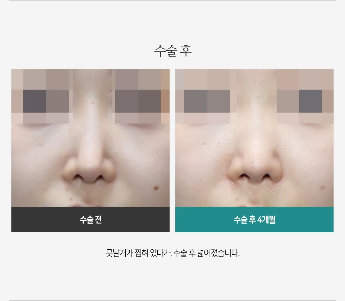
The alae, which had been pinched, widened after surgery.
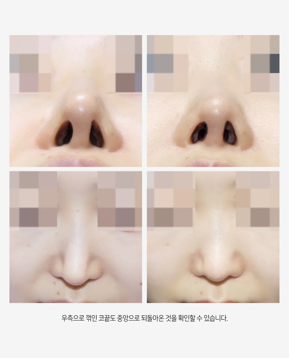
The tip, which had been bent to the right, can also be seen to have returned to the center.

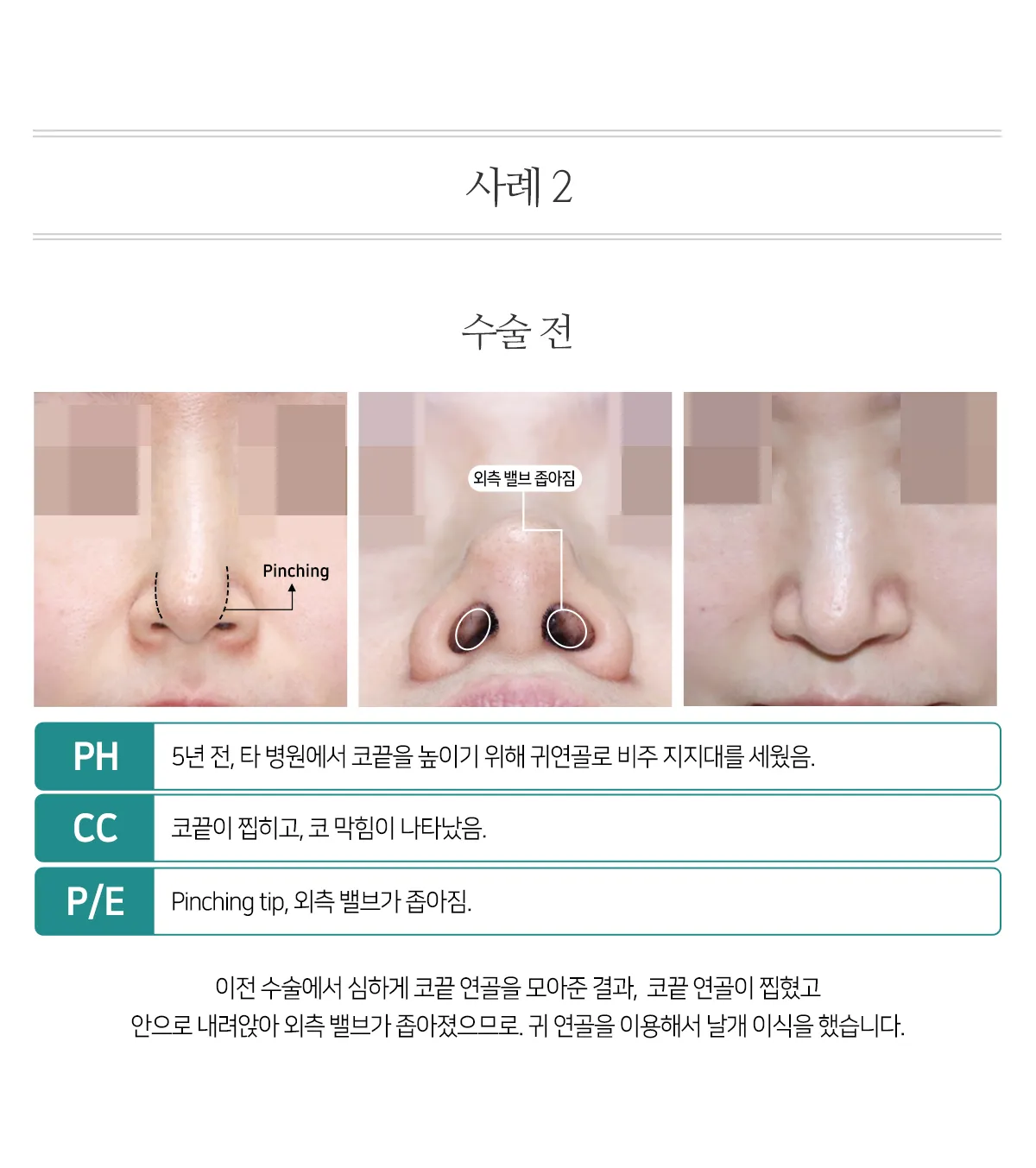
PH
5 years earlier, a columellar strut made of ear cartilage was placed at another clinic to raise the tip.
CC
The tip then became pinched, and nasal obstruction developed.
P/E
Pinched tip and narrowing of the external valve.
Because the tip cartilages had been gathered too tightly in the previous surgery, they had become pinched
and collapsed inward, narrowing the external valve, so a lateral crural strut graft was performed using ear cartilage.
 (A) Intraoperative photo showing the tip cartilages gathered together and pinched.
(B) Ear cartilage harvested and trimmed into a shape suitable for the lateral crural strut graft.
(C) After placement of the lateral crural strut graft.
(A) Intraoperative photo showing the tip cartilages gathered together and pinched.
(B) Ear cartilage harvested and trimmed into a shape suitable for the lateral crural strut graft.
(C) After placement of the lateral crural strut graft.
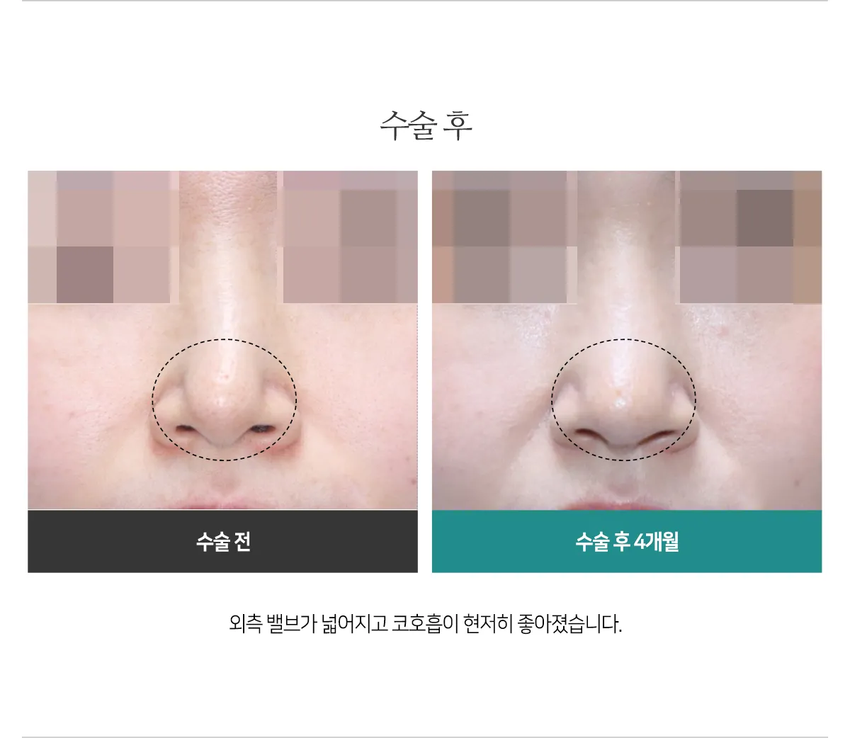
The external valve widened, and nasal breathing improved markedly.

After surgery, the tip cartilages that had collapsed into the nose were lifted, and the external valve widened.
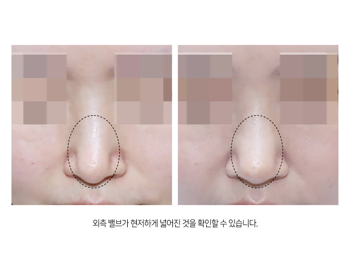
The external valve can be seen to have widened markedly.
 Even though the columellar strut was removed, the height of the bridge and the tip is similar.
Even though the columellar strut was removed, the height of the bridge and the tip is similar.