

The Risks of Diced Cartilage Rhinoplasty #DicedCartilageRhinoplastySideEffects #DicedCartilageRevisionCases

Recently, rhinoplasty that uses cartilage finely diced into small particles
seems to be widely performed and actively promoted.
As if diced cartilage rhinoplasty were opening a new world of plastic surgery.
But just as, in all things, where there is light there is always a dark shadow, the same is true of diced cartilage rhinoplasty,
so following or trusting it unconditionally is not advisable, and its benefits and drawbacks must be weighed.
Today, we will look at the advantages and disadvantages of diced cartilage rhinoplasty, and its risks, through cases.
How Is Diced Cartilage Rhinoplasty Performed?
Recently, more patients have been coming to us after surgery with diced ear cartilage or diced costal cartilage.
Having recognized the problems of silicone implants,
the intention to use autologous cartilage to make up for them is, I think, a good one.
So today I would like to look at diced cartilage rhinoplasty,
beginning with an explanation of the surgical technique.
Rhinoplasty using diced cartilage includes
1) harvesting cartilage, dicing it finely into very small particles,
then gathering and setting the particles in the desired shape before placing them in the nose;
2) wrapping the finely diced cartilage in fascia, absorbable mesh, or a similar material and placing it on the dorsum;
and 3) loading the particles into a syringe and laying them onto the dorsum and tip as if spraying them.
Advantages and Disadvantages of Diced Cartilage Rhinoplasty
Because diced cartilage rhinoplasty uses autologous cartilage, at first glance
it may seem to have few side effects and to be safe,
but in reality it is a technique that also has a number of disadvantages.
Advantages
The surgery is simple and the surgery time is short.
Disadvantages
The length, angle, and other features of the nose cannot be adjusted,
and complex nasal deformities such as contracture, tip deformity, a deviated nose, or an upturned nose
cannot be corrected; it can be used simply to raise the height of the bridge and tip.
Side effects are highly likely to occur.
What Side Effects Does This Technique Have?
It would be helpful to explain the side effects together with their causes.
01. The most common side effect is infection.
02. The particles spread out, and the desired shape is not maintained.
What Is the Solution When Side Effects Occur?
The solution is to remove all of the diced cartilage
and then correct the nose with a method carefully chosen to suit its condition,
but because fine particles are scattered here and there, removal must be meticulous,
and when infection has occurred, the nasal framework and skin are badly damaged, which makes restoration demanding.
Because the skin and soft tissue become thin and damaged, skin irregularities and adhesions can occur, and these must be prepared for in advance.
For the nose to regain a normal shape, the skin thickness must recover sufficiently,
and the condition of the framework of the bridge and tip must be accurately assessed
and a thorough plan drawn up, so considerable experience is required.
Today, we will look at how we operated on patients who came to our clinic because of side effects after surgery with diced cartilage,
and at the results.

PH
20 years earlier: bridge and tip raised with Gore-Tex -> the Gore-Tex implant showed through at the tip
10 years earlier: corrected with ear cartilage because the implant showed through at the tip. (onlay graft)
8 months earlier: autologous diced costal cartilage used to raise the bridge and tip. (open) -> the diced costal cartilage went too far up, as far as the glabella,
making the nose long and straight-lined. A lump-like protrusion formed inside the columella.
1 month earlier: costal cartilage harvested again for revision surgery with diced costal cartilage. (open)
CC
One month earlier: from 9 days after the second diced costal cartilage surgery, the dorsum turned red, with swelling and a heavy feeling.
Yellow fluid came from inside the nose. Irrigated 4 times at the clinic that performed the surgery.
The patient came to our clinic while receiving antibiotic injections.
P/E
The dorsum was red and swollen.
On the columella, there were a small hole and a pus-filled lump caused by infection,
and the columella as a whole was swollen and distended.
DX
Infection
OP
The area around the diced costal cartilage in the dorsum, tip, and columella was red with pus collected,
and the swelling gave it a thickened appearance, findings of infection, so removal surgery was performed.

When we made an incision and looked inside, pus and exudate had collected around the diced costal cartilage laid on the dorsum and tip,
and the diced costal cartilage had failed to keep a consistent shape and was scattered, so it had no aesthetic effect.
On the dorsum, a piece of Gore-Tex remained in the layer beneath the diced costal cartilage, firmly attached to the framework;
this was left behind because the Gore-Tex from the earlier augmentation had not been completely removed,
and this piece of Gore-Tex is thought to be one of the factors that caused the infection.
Dissection through the columella showed costal cartilage positioned as a septal extension graft or extended spreader graft,
so all of it was removed.

The piece of Gore-Tex is thought to be one of the main causes of the surrounding infection,
and cartilage in particle form is vulnerable to infection, which spreads easily

Before removal, the dorsum was red and swollen, and yellow pus had collected at the tip.
The dorsum and tip looked broad and blunt.
Although the diced costal cartilage is said to be gathered into a fixed shape with clotted blood or artificial adhesive (glue) before placement,
the pressure of the skin makes it hard for the diced costal cartilage to keep a consistent shape, which is why the contour of the nose was broad.
In addition, when diced costal cartilage is contaminated with bacteria, its large contact area and dispersed form make it vulnerable to infection.
At 3 weeks after removal, the skin of the dorsum was uneven, and deformity of the tip and columella had appeared.
Diced costal cartilage is difficult to remove through a closed approach,
and because the particles are as finely divided as grains of sand, removal is demanding and is likely to damage the soft tissue.
When infection is present, erosion of the skin is even more severe and leads to damage.
As a result, the skin becomes uneven, and during the contraction period severe deformity can appear not only in the skin but also in the tip cartilages.

Before Removal
The infection was severe, with pus collected, and the entire tip was red and swollen.
After Removal
Marked irregularities appeared because of damage to and deformation of the cartilage and skin.

Before Removal
The glabella was high and looked unnatural, the dorsum was uneven, and the dorsal contour was broad
After Removal
Contraction caused the columella to sink in, and the tip was notched
Distinct irregularities remained because of skin damage

After removal, columellar depression, skin irregularities on the dorsum, pigmentation, and tip deformity remained.
Secondary surgery will be needed in 6-12 months, once skin regeneration has matured,
and the patient is waiting while receiving autologous blood-derived stem cell injections.

PH
1st. 10 years earlier, at another clinic: silicone + ear cartilage to the tip (open)
-> The skin thinned, and the tip became pointed and red.
2nd. 6 years earlier, at another clinic: autologous diced costal cartilage + septal cartilage used at the tip.
CC
The dorsum was uneven. The bridge was thin and hard, the nostrils were uneven, and the nostrils were pointed and pinched,
The tip was red. Tightness. A feeling of blood pooling
P/E
The dorsum and tip were hard and showed through because of the diced costal cartilage. The tip was pointed and red.
The bridge was crooked.
OP
Removal of the diced costal cartilage, trimming of the SEG tip, and alar cartilage repositioning

Before Surgery
The bridge was crooked, and the outline of the straight-line diced costal cartilage showed through and was visible,
and the nostrils were pulled up into a point.
After Surgery
The dorsum became natural, and nothing shows through any longer,
and the pointed, pinched tip came down and softened.
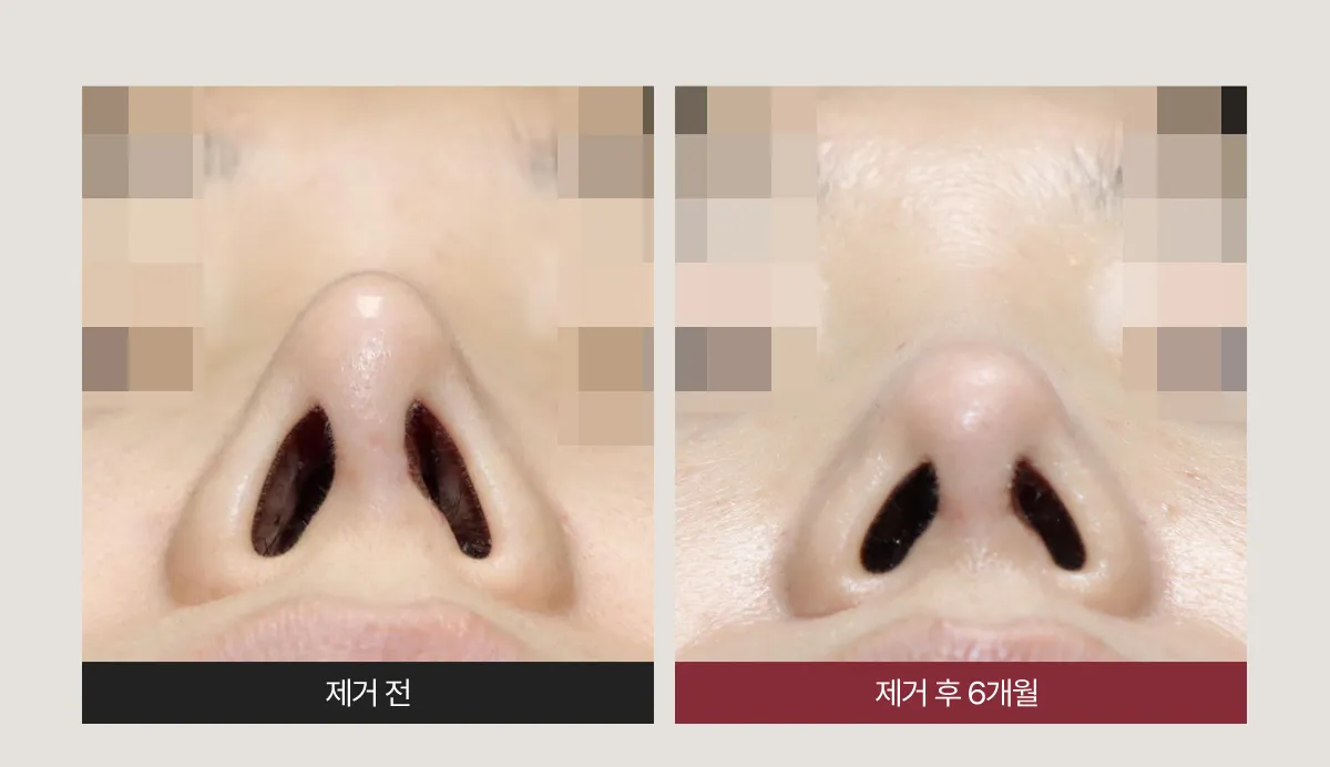
The pointed tip and nostrils became fuller and softer after surgery.

The tilted, pointed tip was centered and softened after surgery.

There was a step just above the tip that made the profile look bent, and it became straight after surgery.
The upturned tip also came down naturally, and the sharp bridge and tip became natural.

The irregularity above the tip disappeared, and the profile became straight and natural.

PH
2020.5: 3 types of rhinitis surgery, augmentation with silicone, and donor costal cartilage for the tip.
Spreader graft with mesh, osteotomy. Alar reduction (lateral incision), open approach -> inflammation and swelling
1 month later, only the silicone was removed -> the bridge became lower
2020.12: bridge augmentation by diced costal cartilage injection (closed) -> completely resorbed
2021.10: augmentation with donor costal cartilage (open)
CC
The tip was crooked, the nose often swelled and reddened from an inflammatory reaction, and the dorsum was broad.
OP
2024.4.11: removal of the septal SEG and spreader mesh + removal of inflamed tissue + partial removal of the diced costal cartilage
+ removal of the donor costal cartilage (the septal cartilage had dissolved)
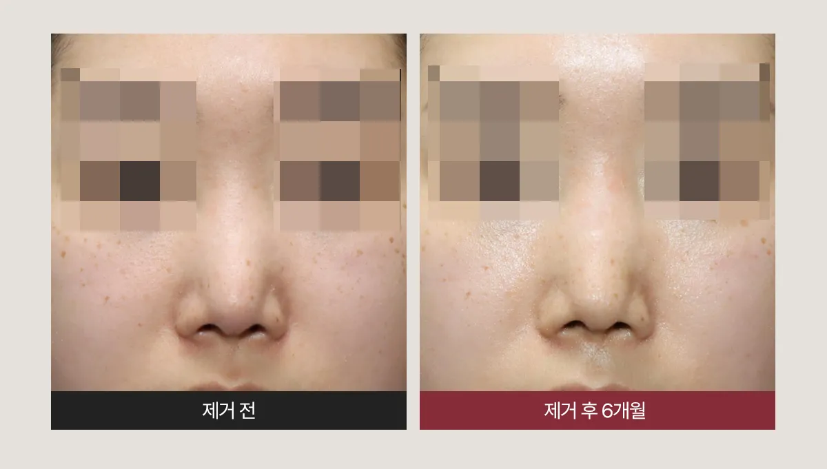
After the removal surgery, the inflammatory reaction of redness and frequent swelling disappeared.

Even with removal alone, the nose became longer and slimmer.

The dorsal line now runs straight and looks natural.

PH
August 2019, at another clinic: bridge augmentation with donor diced costal cartilage + SMR + turbinectomy
+ columellar strut with septal cartilage (open) -> resorption at the bridge
March 2020, at another clinic: additional grafting to the bridge with donor diced costal cartilage.
CC
Resorption left the glabella and the middle of the nose sunken, so there was little difference compared with before surgery, and the patient wanted removal.
OP
Complete removal of the donor diced costal cartilage on the dorsum, the septal extension graft, and the donor diced costal cartilage at the tip & tip cartilage repositioning.
** Because there was no septum, part of the extension graft was left in place. Because the columellar strut cartilage had weakened,
the septal cartilage graft at the tip was removed and used to reinforce the septum. Cartilage graft to the left external nasal valve; Silastic sheet (+)
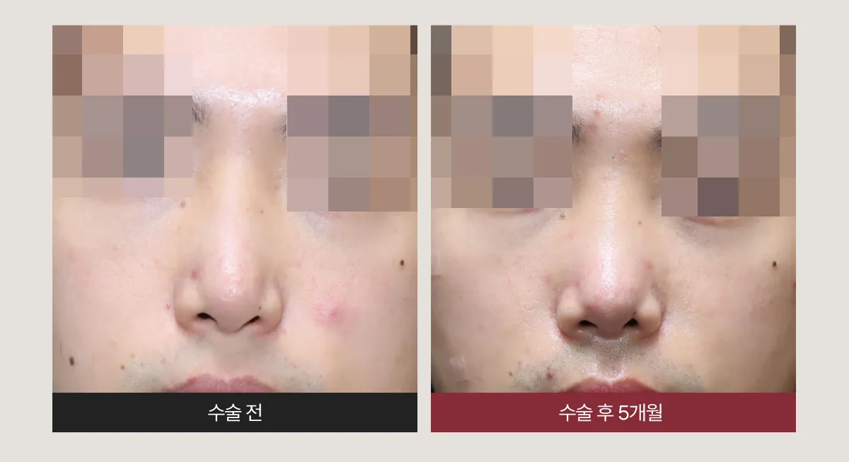
After removal, the high glabella returned to normal and the shape of the nose became more natural.

The crooked columella and nostrils became straight after surgery.
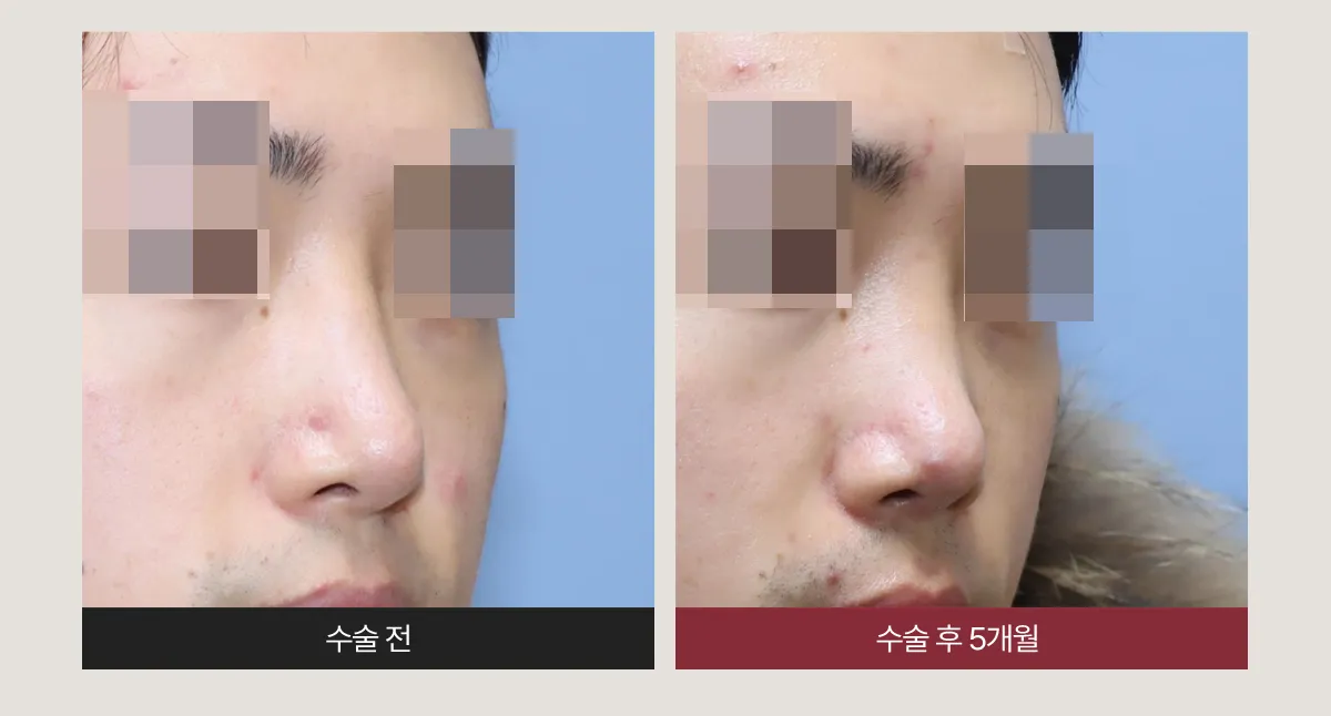
After the diced costal cartilage at the glabella was removed, the dorsal line became natural.

After surgery, the length of the nose and the dorsal line look natural.