

Severe Infection After Silicone Rhinoplasty / A Case of Implant and Infected Tissue Removal

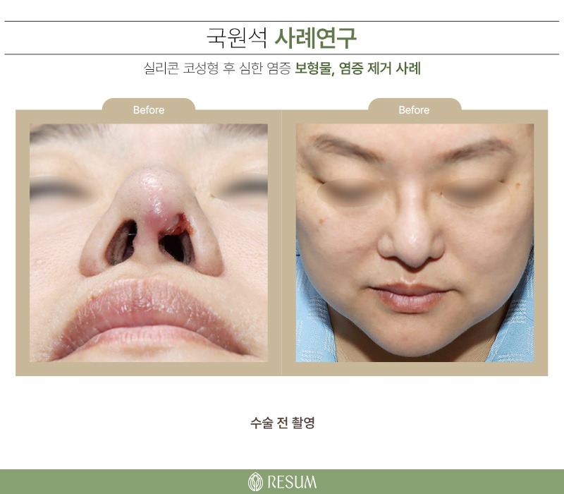
Woman in her 40s
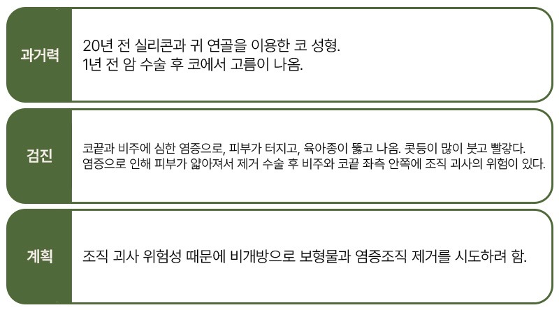
History : Rhinoplasty with silicone and ear cartilage 20 years ago. Pus began draining from the nose after cancer surgery 1 year ago.
Examination : Severe infection of the tip and columella, with the skin broken open and a granuloma protruding through it. The dorsum is markedly swollen and red.
Because the infection has thinned the skin, there is a risk of tissue necrosis at the columella and the inner left side of the tip after removal surgery.
Plan : Because of the risk of tissue necrosis, removal of the implant and infected tissue was to be attempted through a closed approach.
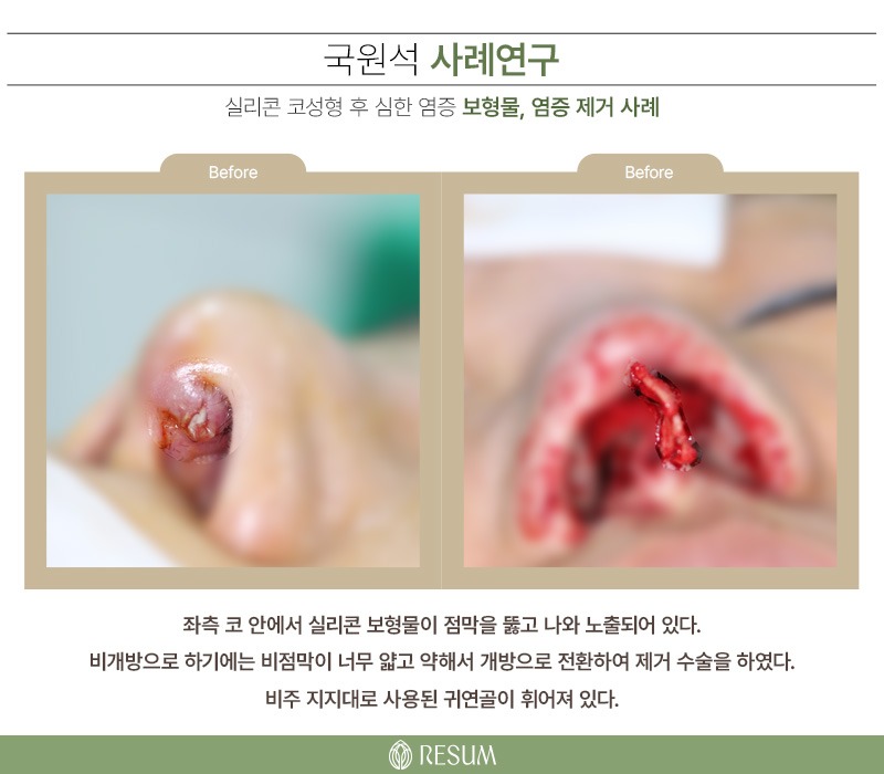
Inside the left side of the nose, the silicone implant had broken through the mucosa and was exposed.
The nasal mucosa was too thin and fragile for a closed approach, so the removal surgery was converted to an open approach.
The ear cartilage used as a columellar strut was bent.
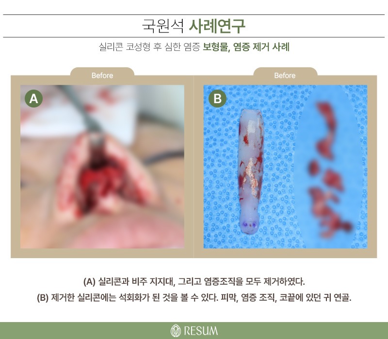
(A) The silicone, the columellar strut, and the infected tissue were all removed.
(B) Calcification can be seen on the removed silicone. Capsule, infected tissue, and the ear cartilage that had been at the tip.
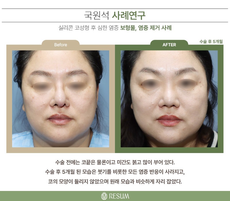
Before surgery, not only the tip but also the glabella was red and markedly swollen.
At 5 months after surgery, the swelling and all other inflammatory responses have resolved,
the nose has not turned upward and has settled into a shape close to its original appearance.
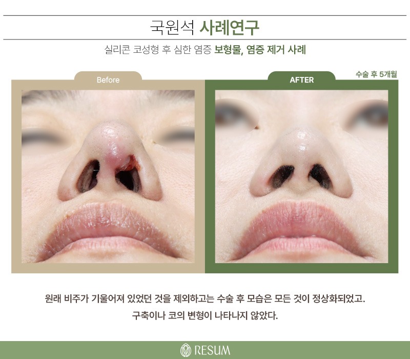
Apart from the columella, which had been tilted originally, everything has normalized after surgery,
and no contracture or nasal deformity has appeared.
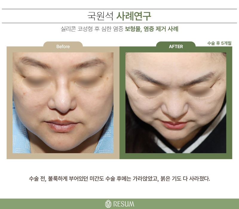
The glabella, which bulged with swelling before surgery, has also subsided after surgery, and the redness is entirely gone.
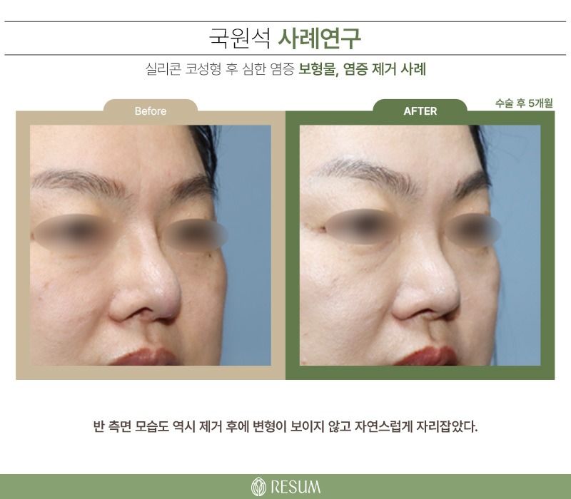
In the three-quarter view as well, no deformity is seen after removal, and the nose has settled naturally.
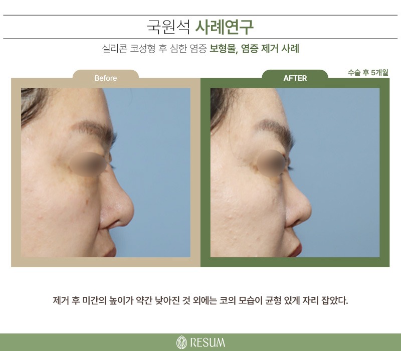
Apart from a slight decrease in the height of the glabella after removal, the nose has settled into a balanced shape.